Multifocal atrial tachycardia (MAT) is an irregularly irregular rhythm, typically between 100-150 beats per minute, that arises from multiple ectopic foci within the atria. It is a rare condition that is most often seen in patients with advanced lung disease, such as chronic obstructive pulmonary disease. MAT is defined by a rate > 100 beats per minute with at least three morphologically distinct atrial complexes, varying P-P intervals, and an isoelectric baseline between P waves. The mechanism is not completely understood but is believed to be caused by either reentrant centers, increased atrial automaticity, or triggered centers; and is often associated with respiratory failure. Underlying hypoxia or hypercarbia, right atrial dilation, and increased sympathetic drive are typical physiologic stressors contributing to development of MAT in respiratory failure. Clinicians should focus on treatment of the underlying cause in order to resolve the MAT. Unfortunately, the development of MAT during an acute illness/exacerbation should be viewed as a poor prognostic indicator, with a significant in-hospital mortality associated with it during acute illness.
Multifocal Atrial Tachycardia

Examples
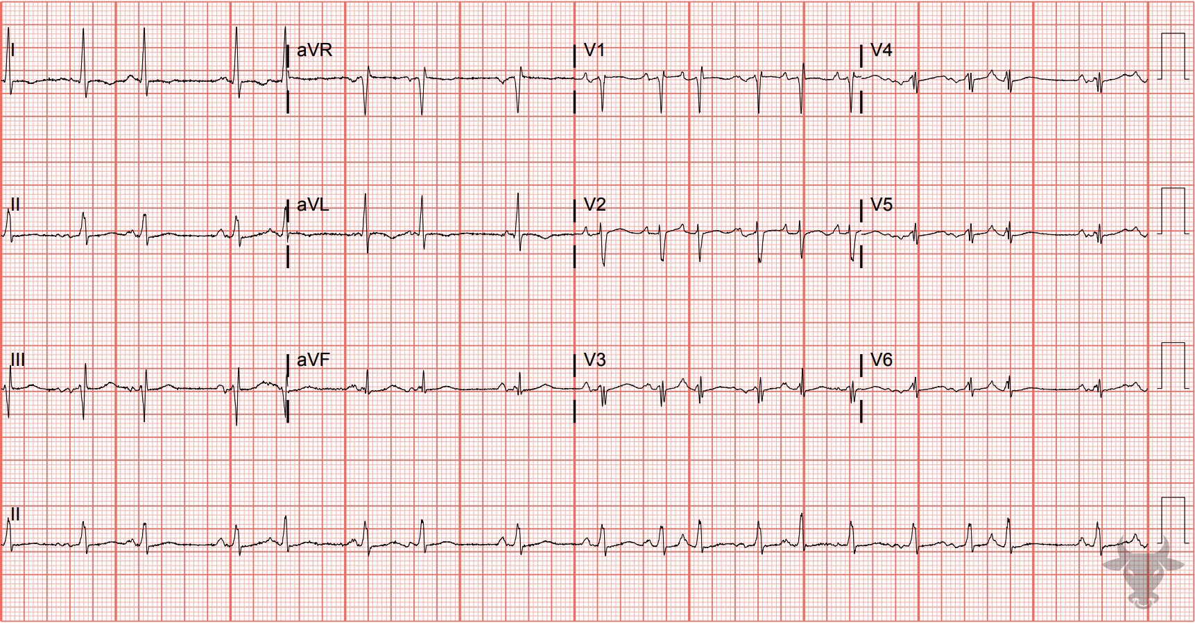
Multifocal Atrial Tachycardia
Multifocal atrial tachycardia in a patient with chronic obstructive lung disease. There is also low voltage due to increased impedance from hyperinflated lungs.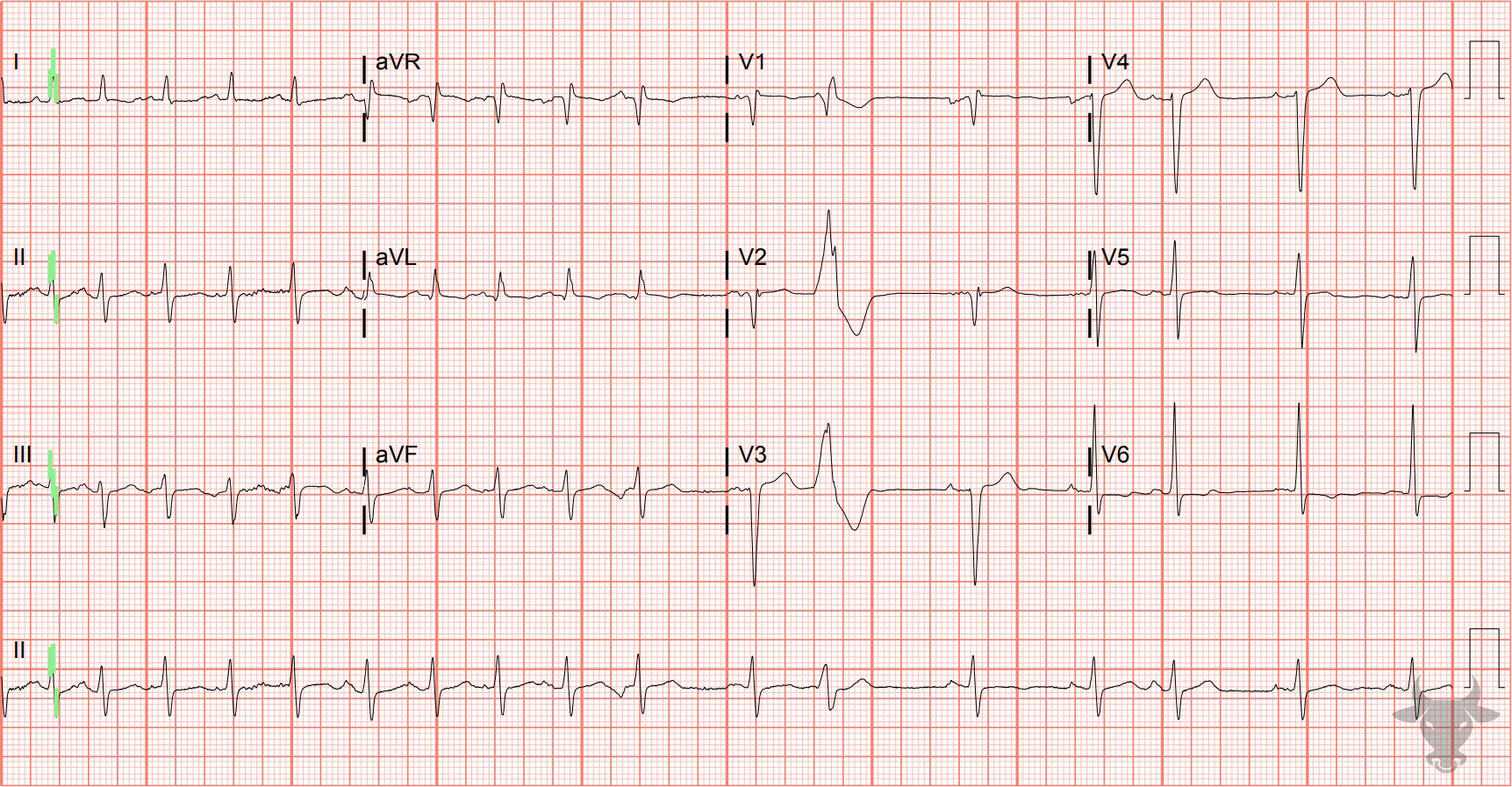
Multifocal Atrial Tachycardia
Multifocal atrial tachycardia in a patient with heart failure and chronic obstructive pulmonary disease.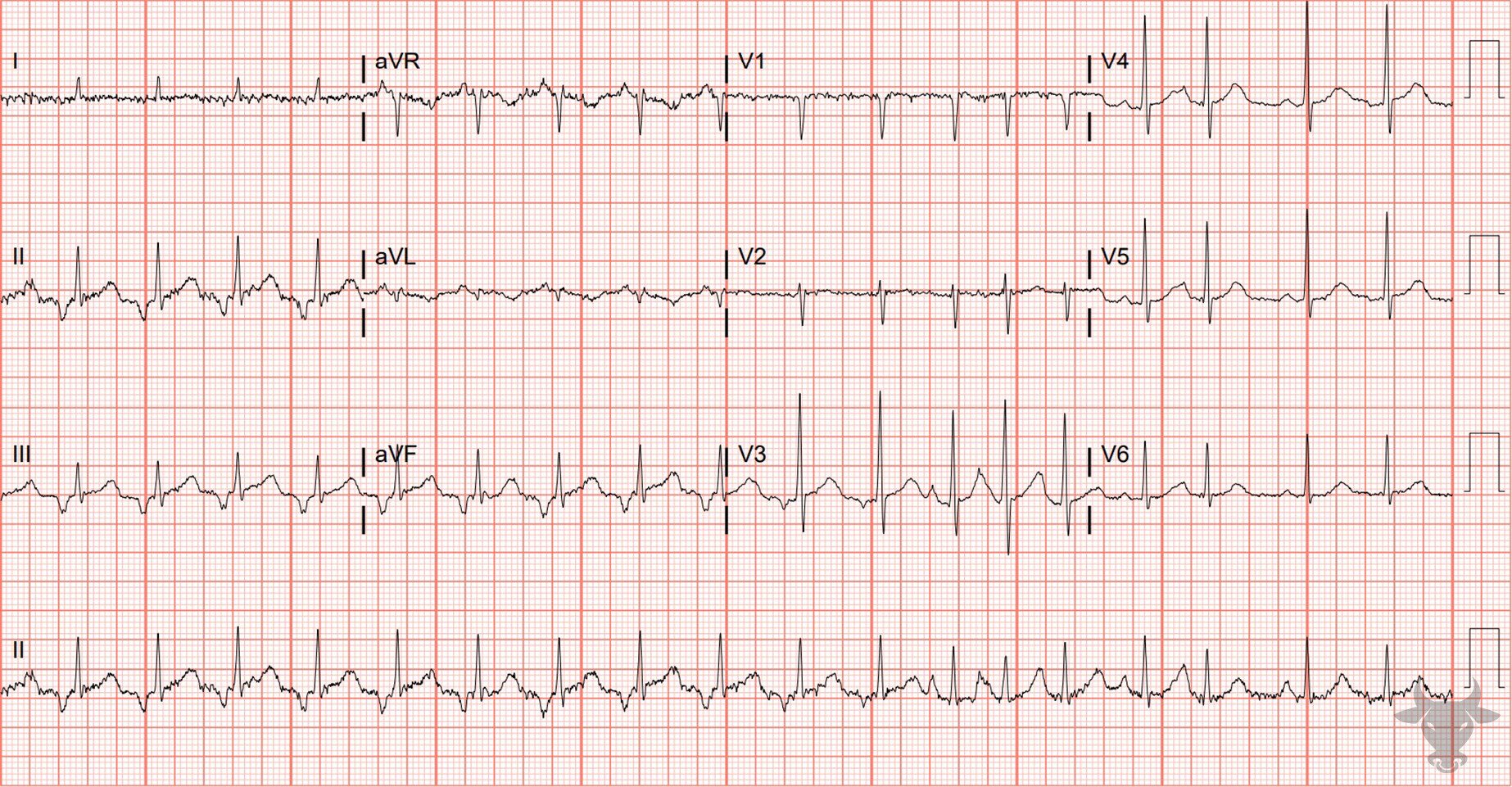
Multifocal Atrial Tachycardia
The presence of three different P wave morphologies confirms the diagnosis in this patient with chronic obstructive lung disease.References
- Wagner GS, Strauss DG. Marriott’s Practical Electrocardiography. 12th ed. Lippincott Williams & Wilkins; 2014
- Surawicz B, Knilans TK. Chou’s Electrocardiography in Clinical Practice. 6th ed. Elsevier; 2008.