The traditional definition of low voltage is QRS amplitudes less than 5 mm in all limb leads or less than 10 mm in all precordial leads. The differential for low voltage can be conceptualized by considering two categories: cardiac abnormalities resulting in diminished impulse generation and increased impedance related to attenuating substances between the heart and surface leads (fluid, air, or adipose). Cardiac causes include prior infarcts, infiltrative cardiomyopathies like amyloidosis, and hypothyroidism. Extracardiac causes include pericardial processes like pericardial effusion or constrictive calcific pericarditis, chronic obstructive pulmonary disease, pleural effusion, subcutaneous or mediastinal emphysema, and obesity. The combination of low voltage and bradycardia should raise the concern for hypothyroidism/myxedema.
Low Voltage

Examples
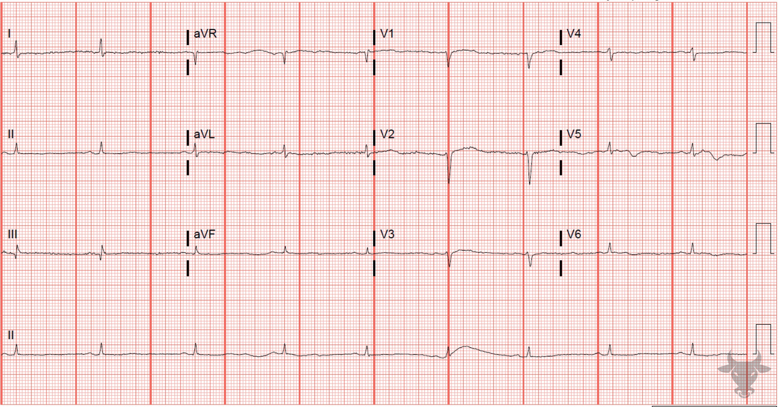
Low Voltage
Low voltage resulting from the decreased impulse generation of myxedema.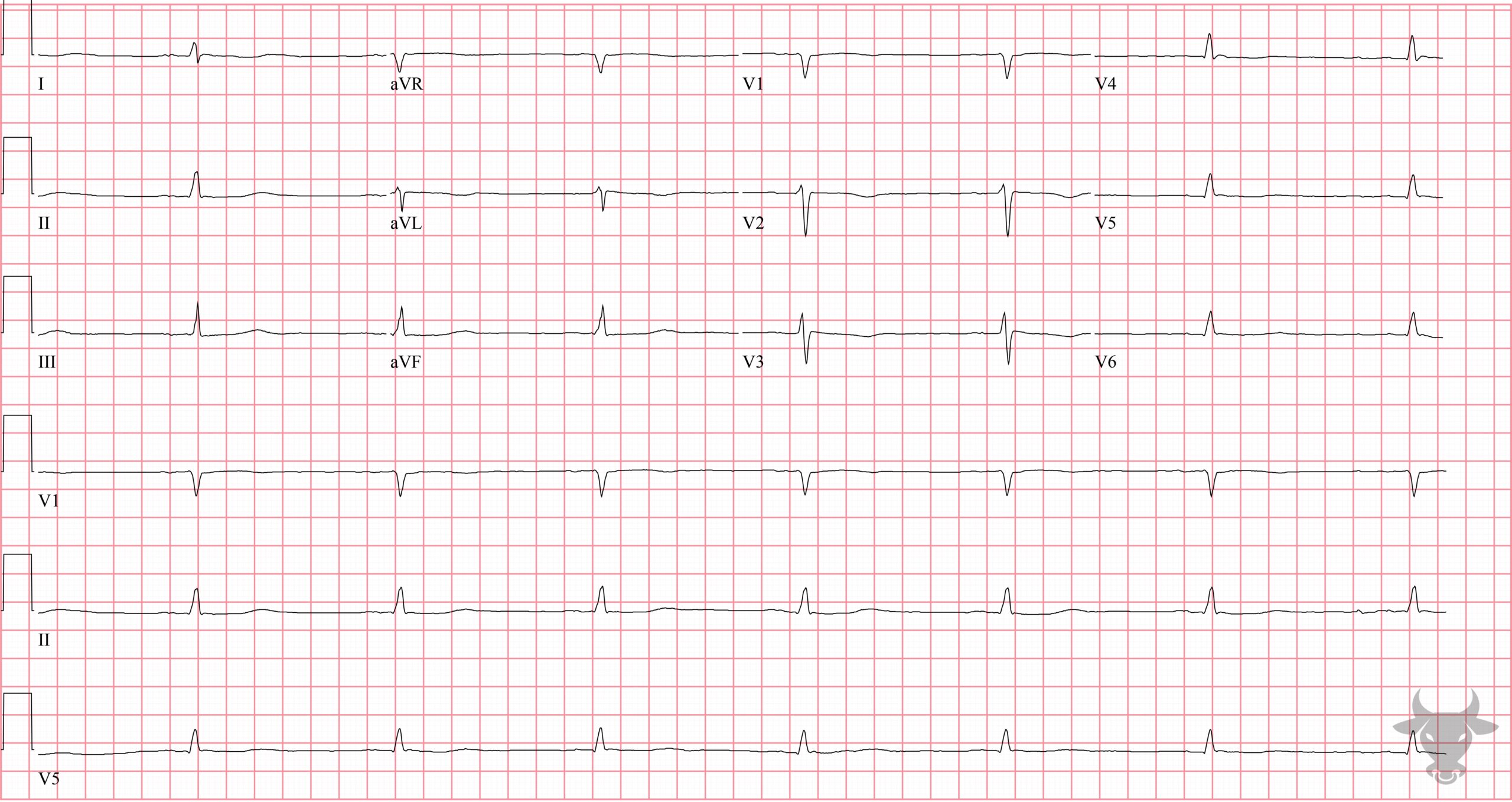
Low Voltage
Bradycardia, prolonged QT, and low voltage all due to myxedema. This patient had a large pericardial effusion. hypothyroidism can cause low voltage through two mechanisms: the direct effects of hormonal deficiency on the generation of cardiac action potentials and via the presence of a pericardial effusion, seen in up to one third of patients with hypothyroidism and, rarely, leading to tamponade. Patients with tamponade or large effusions due to hypothyroidism will characteristically lack a compensatory tachycardic response.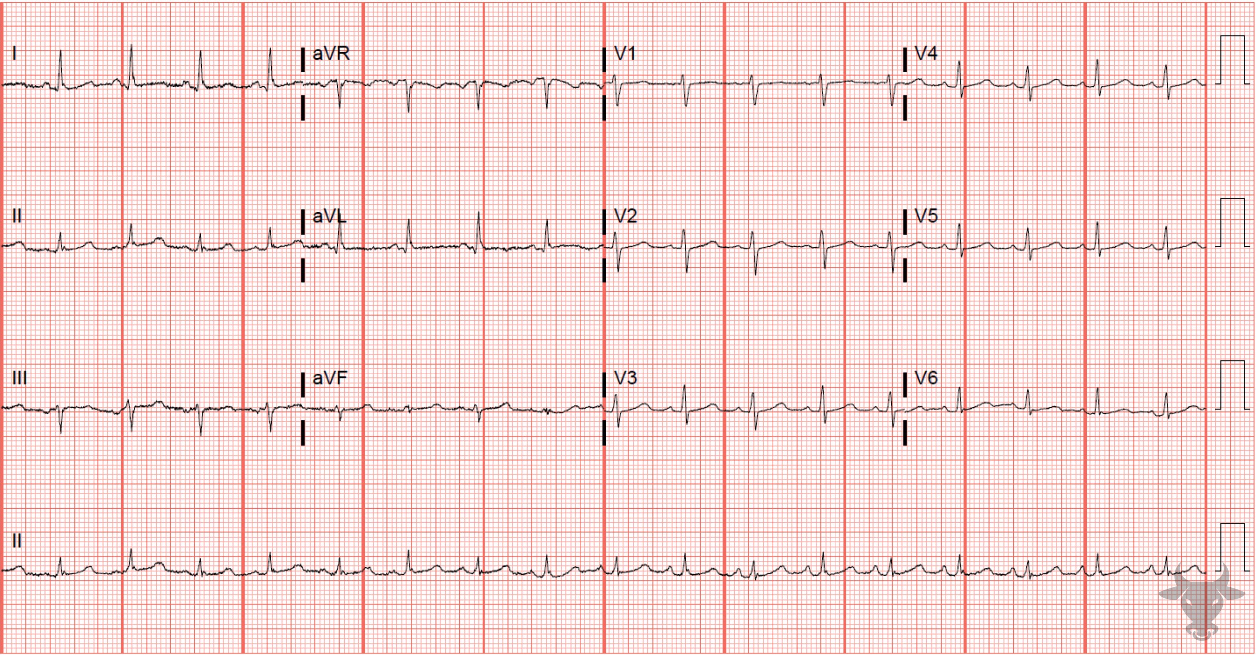
Low Voltage
The QRS amplitudes in the rhythm strip of lead II can be seen to alternate between larger and smaller complexes, a phenomenon known as electrical alternans. Electrical alternans often results from the pendulous-like swinging of the heart inside a fluid-filled pericardium, as with this case.
Multifocal Atrial Tachycardia
Multifocal atrial tachycardia in a patient with chronic obstructive lung disease. There is also low voltage due to increased impedance from hyperinflated lungs.References
- Danzi S, Klein I. Thyroid disease and the cardiovascular system. Endocrinology and Metabolism Clinics of North America. 2014;43(2):517-528.
- Madias JE. Low QRS voltage and its causes. Journal of Electrocardiology. 2008;41(6):498-500.
- Ingram D, Strecker-McGraw MK. Electrical Alternans. StatPearls. Published online on August 11, 2021. Accessed January 3, 2022.