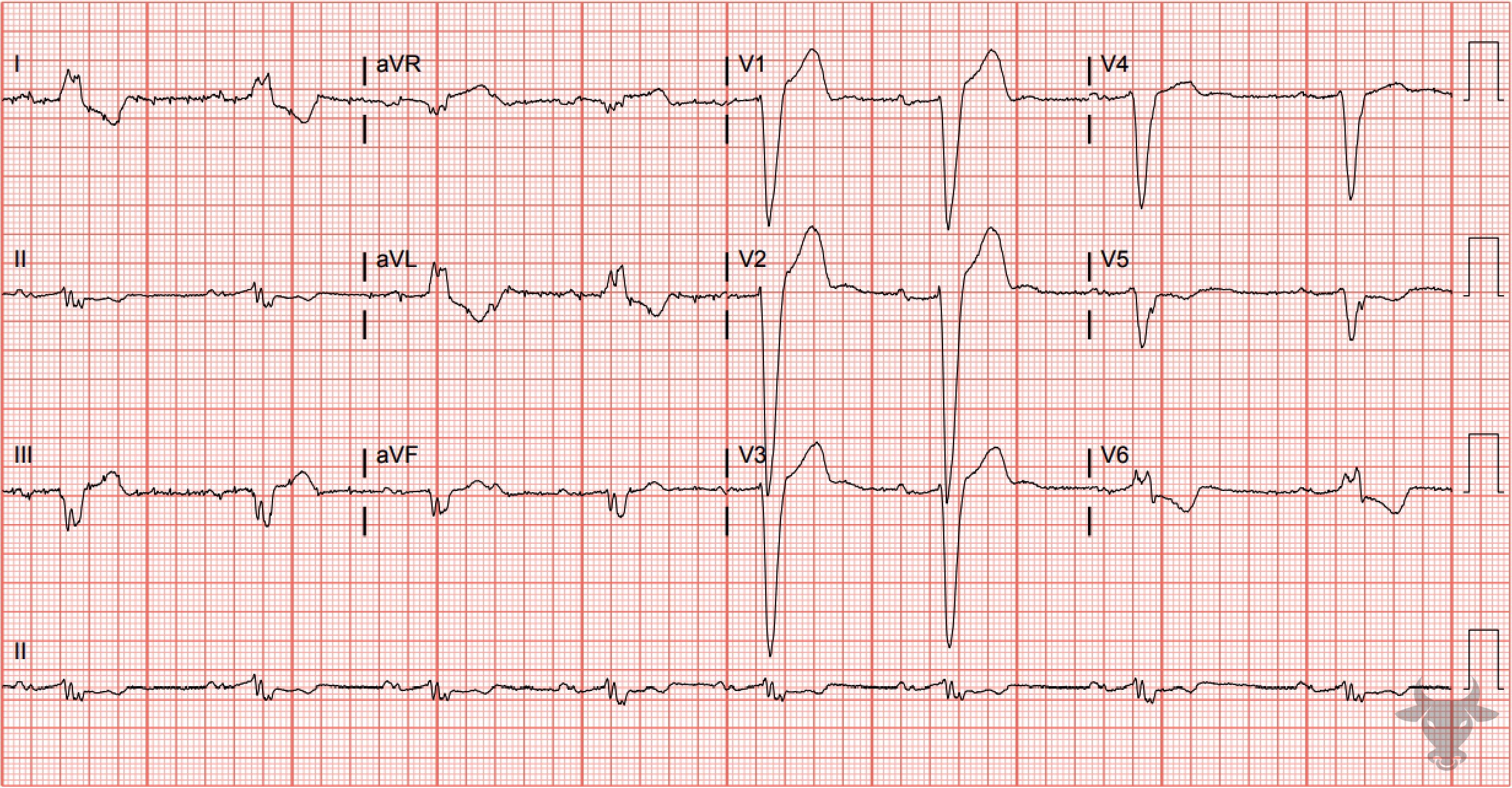
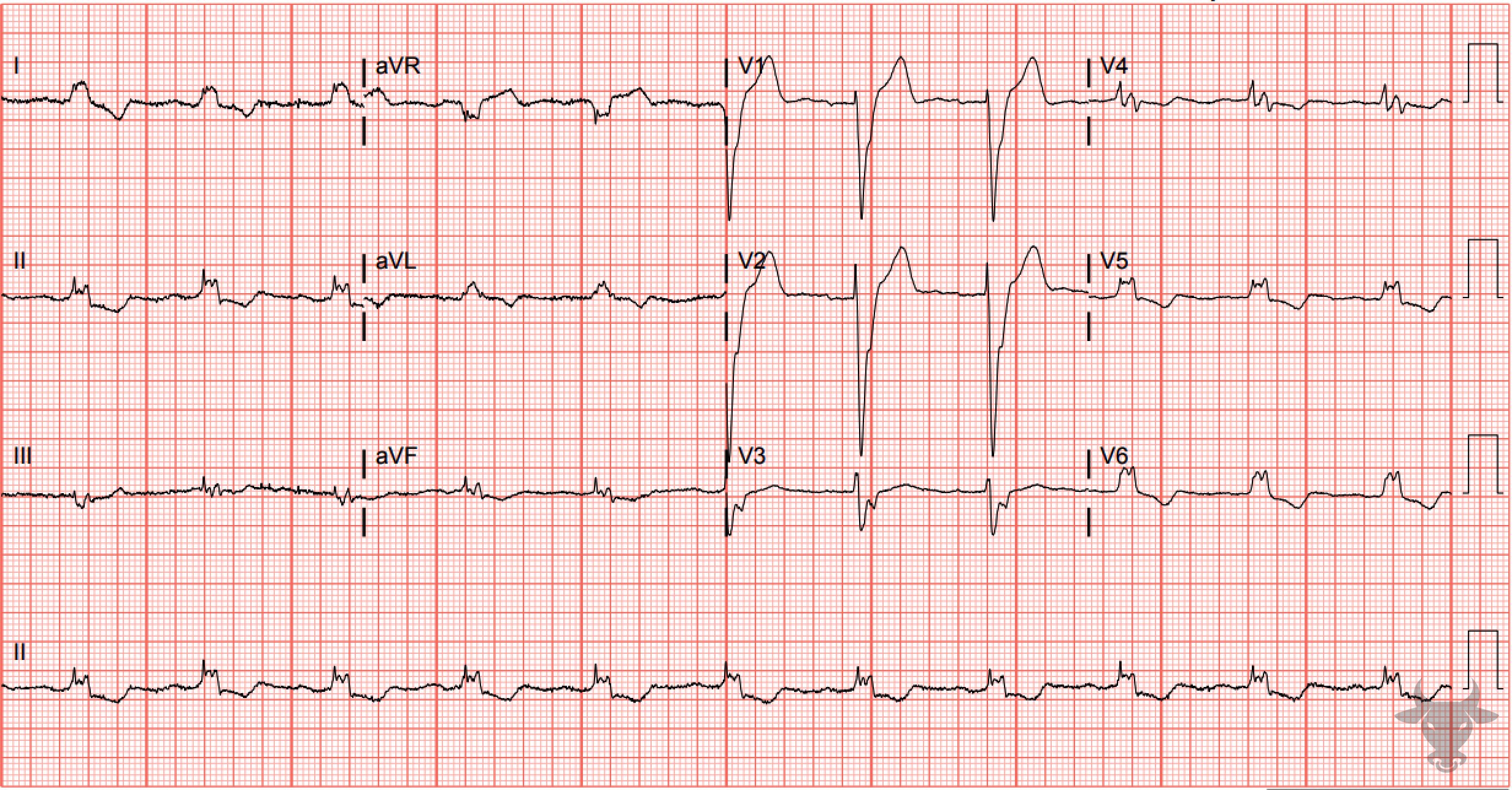
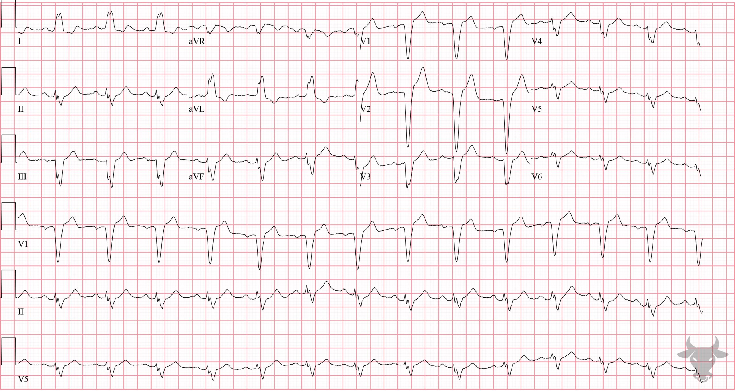
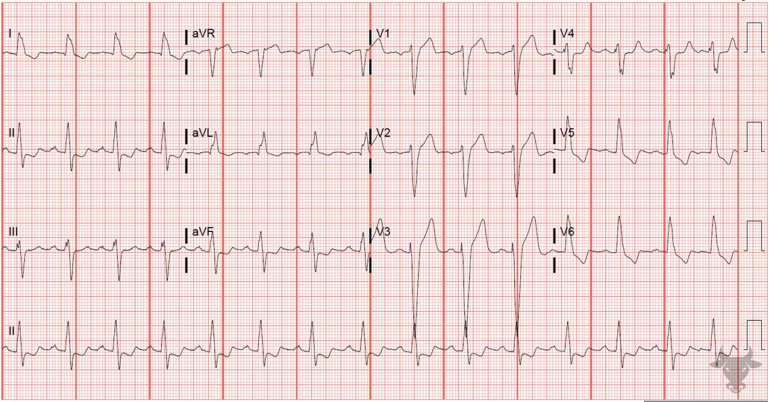
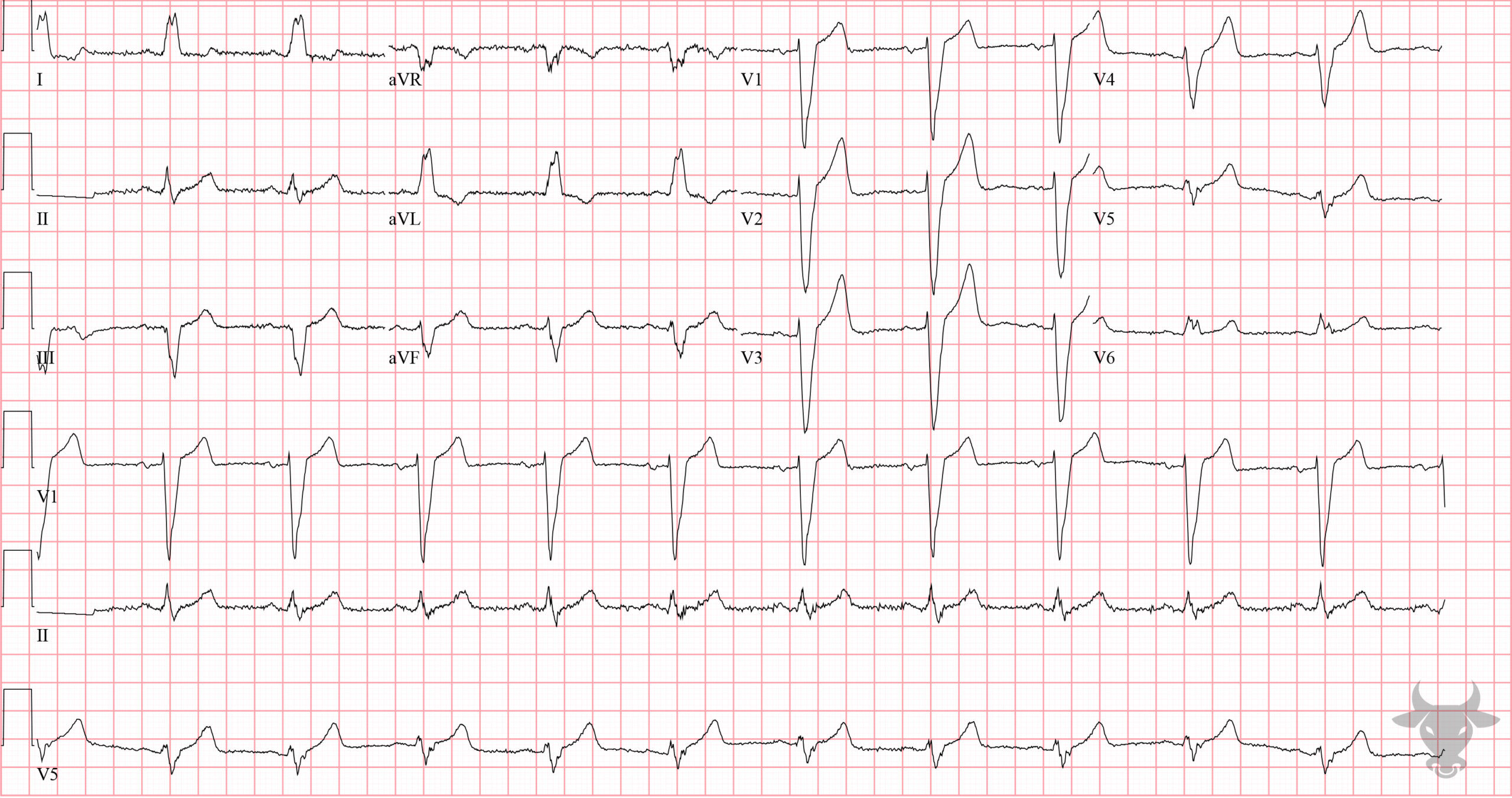
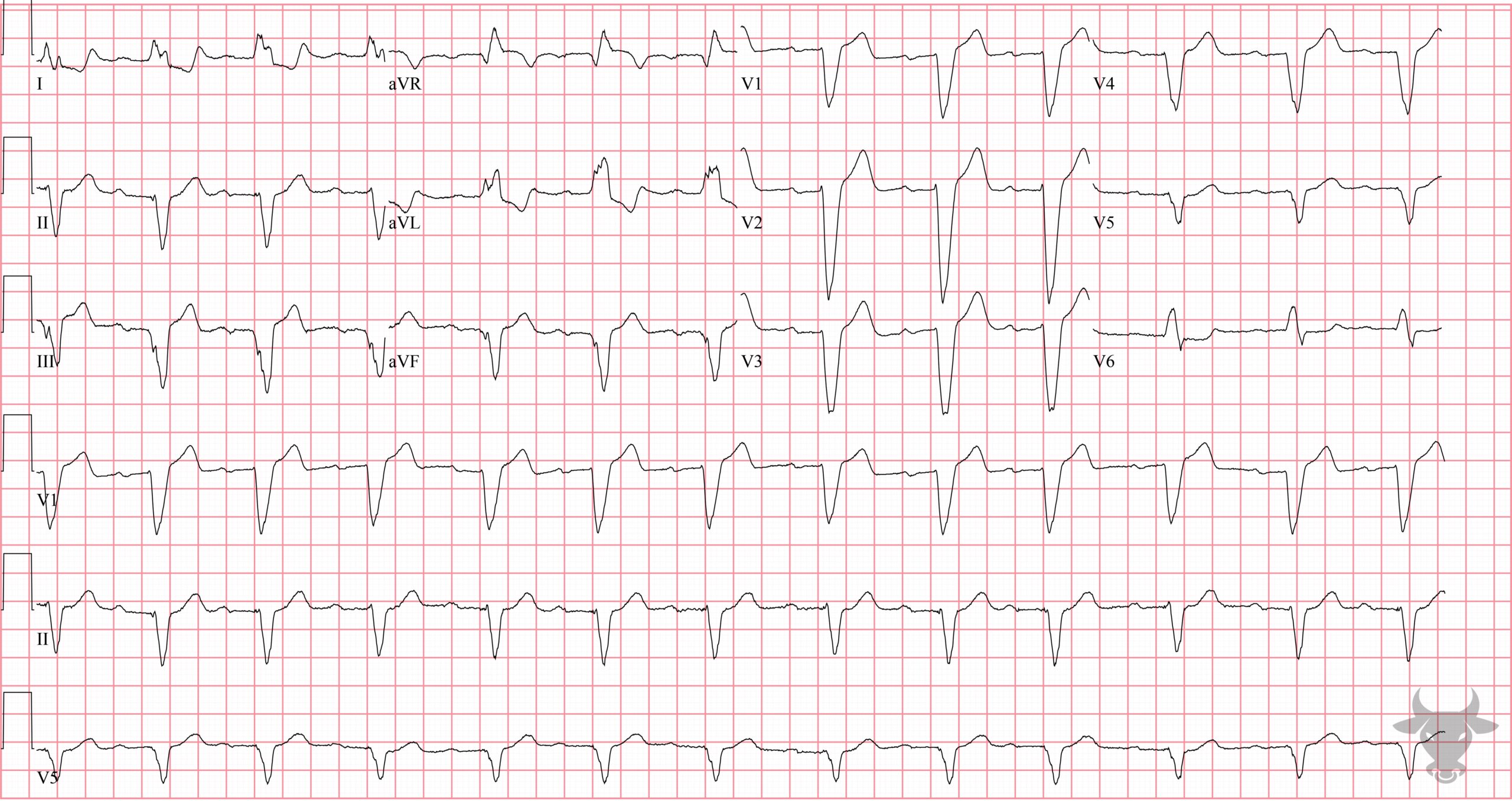
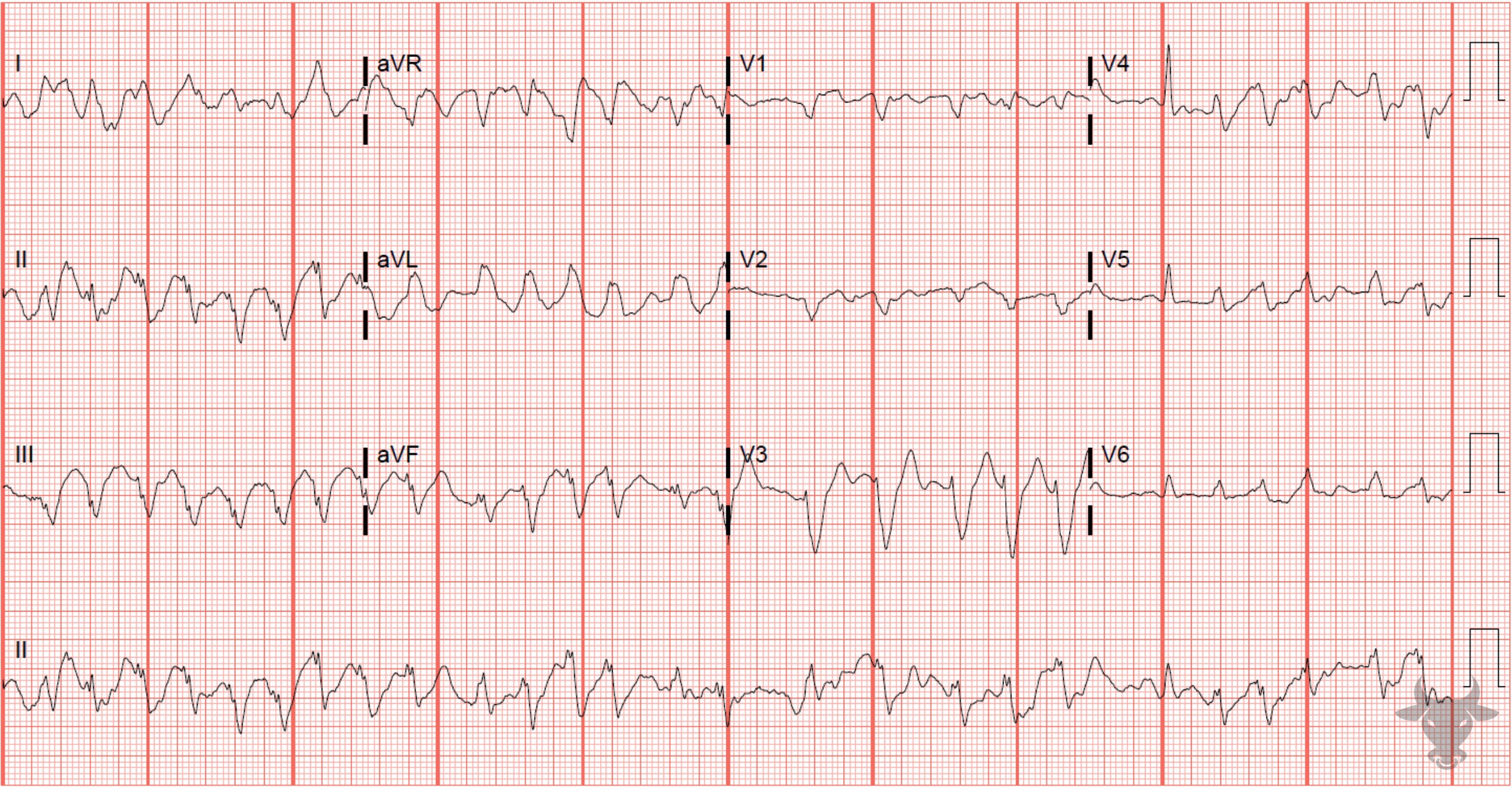
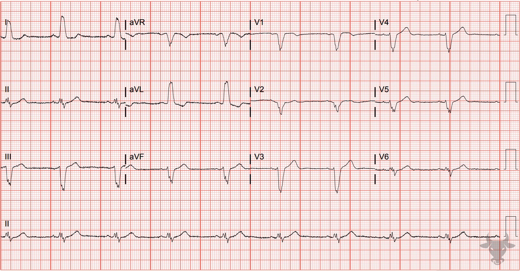
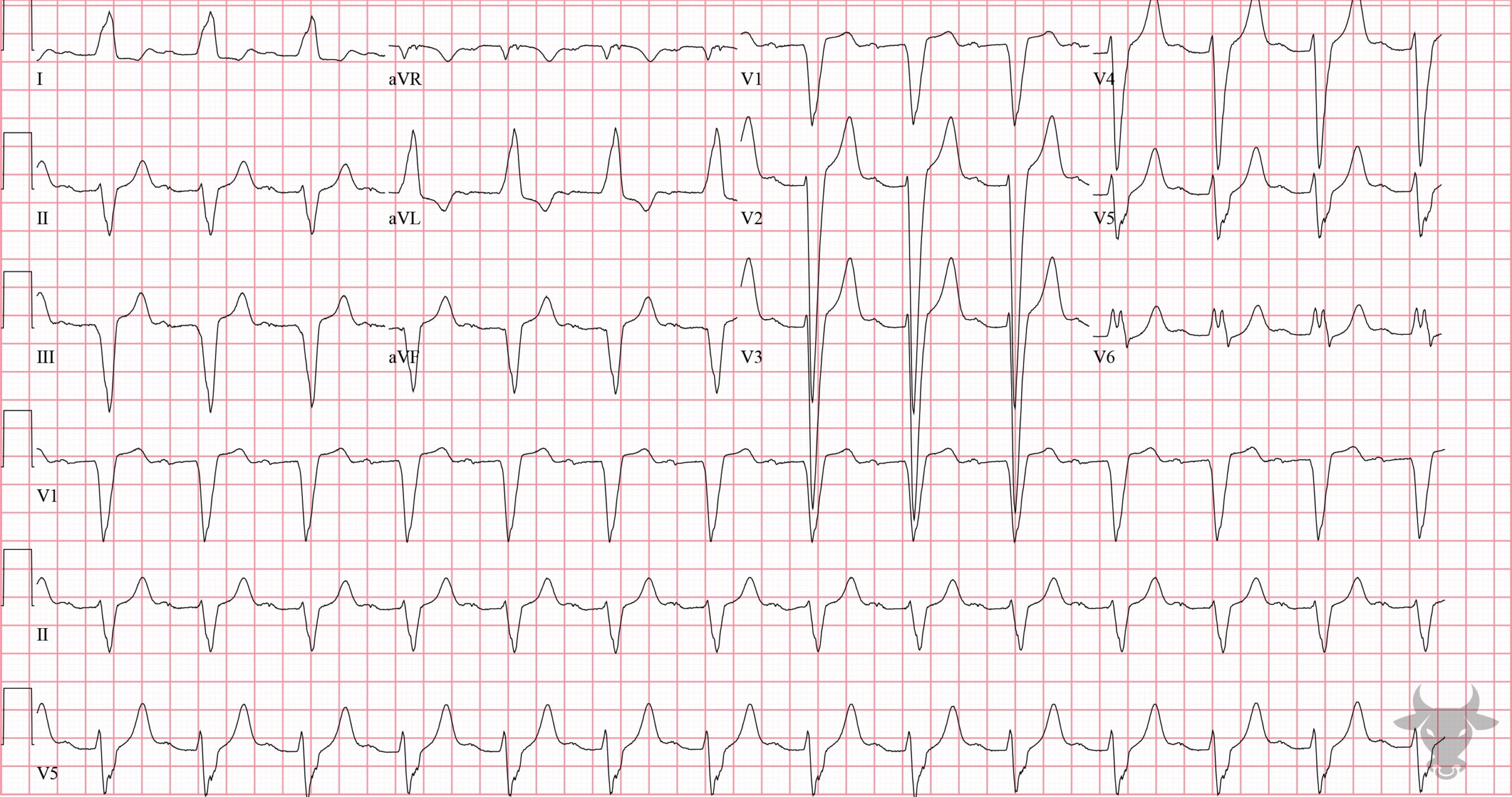
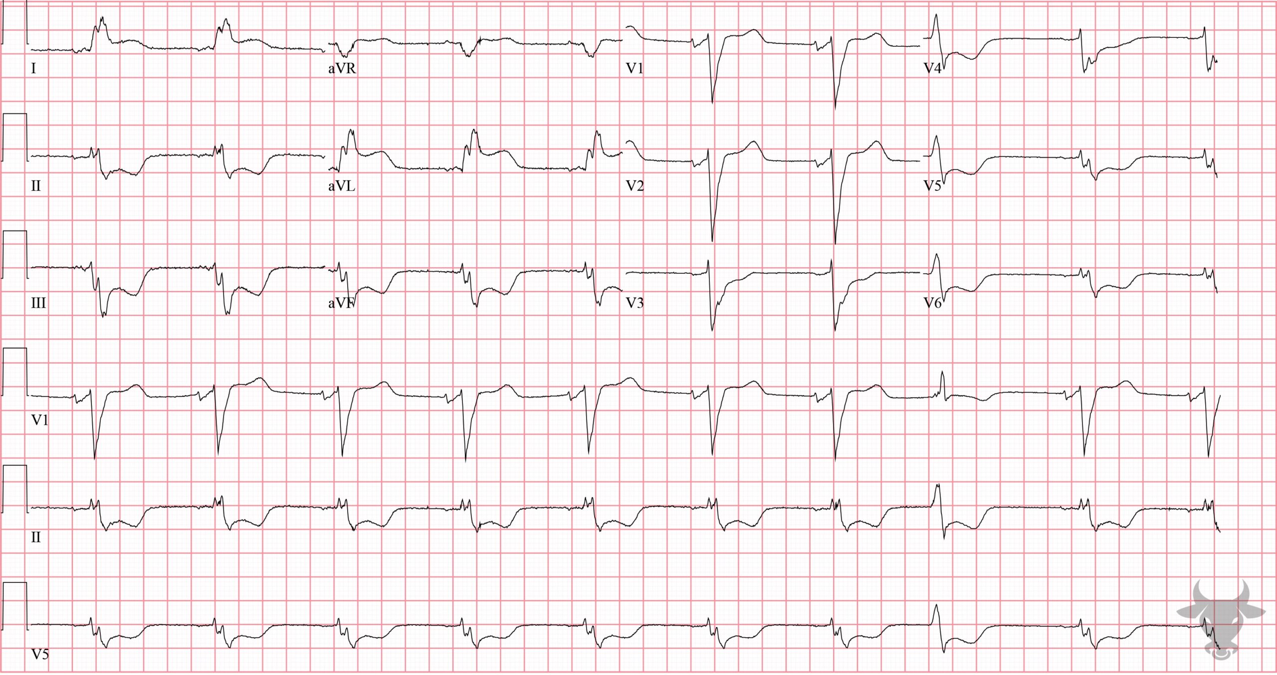
Left bundle branch block (LBBB) occurs when the left bundle no longer conducts, and the signal must pass to the left ventricle via myocyte-to-myocyte conduction. This pattern of conduction is slower than via the specialized conduction system, and results in a wide QRS complex (>120 ms). Conduction disturbances, like bundle branch blocks, result from structural abnormalities of the His-Purkinje system caused by necrosis, fibrosis, calcification, infiltrative disease, electrolyte disturbances, or impaired vascular supply. When conduction is impaired to both left ventricular terminals (the left anterior and posterior fascicles), the result is a LBBB. The criteria are:
- QRS >120 ms
- Dominant S wave in V1
- Broad monophasic or notched R wave in lateral leads (I, aVL, V5, V6)
- +/- left axis deviation
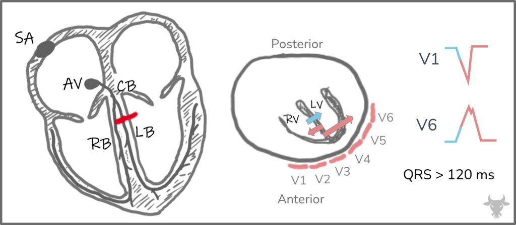
Depiction of depolarization in left bundle branch block. The septum first depolarizes from right to left (blue arrow), followed by near simultaneous depolarization of the right and left ventricles (pink arrows). The resultant vector leads to a negative deflection in V1 and a positive deflection in V6. SA, sinoatrial node; AV, atrioventricular node; RB, right bundle; LB, left bundle; CB, common bundle; RV, right ventricle; LV, left ventricle; red dash represents a block of the left bundle.