Hypokalemia, hypomagnesemia, and hypocalcemia can all cause a prolongation of the QT interval. While hypokalemia and hypomagnesemia both delay the repolarization phase (phase 3) of the of the cardiac action potential (creating wide-based T waves, U waves, or a fusion of both), hypocalcemia prolongs the QT interval by way of extending the plateau phase (phase 2) of the cardiac action potential (lengthening the ST segment but with a normal T wave).
Hypocalcemia

Examples
Hypocalcemia
QT prolongation by way of lengthened ST segment is specific for hypocalcemia (as opposed to broadening of the T wave or T-U fusion). The peaked T waves also suggest hyperkalemia. This is a classic ECG for patients with end-stage renal disease.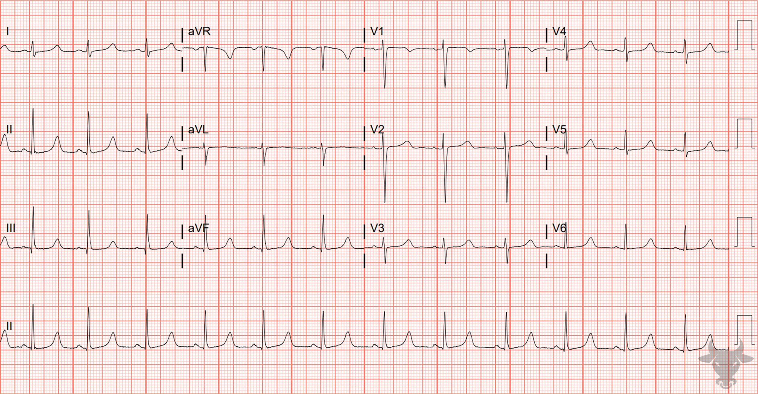
Hypocalcemia
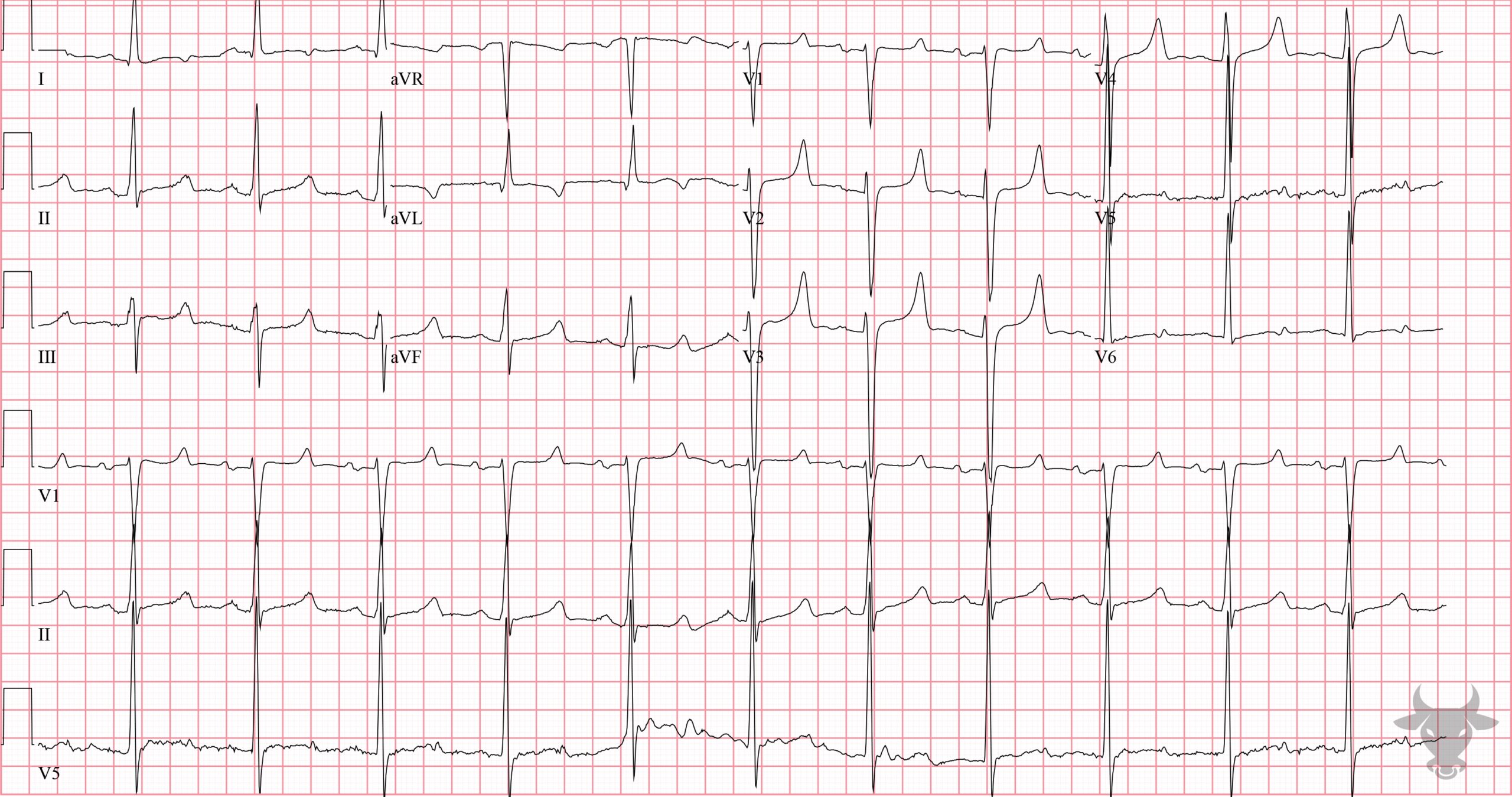
This patient presented with fatigue and was found to have newly discovered renal failure. The calcium was 5.6 mg/dL.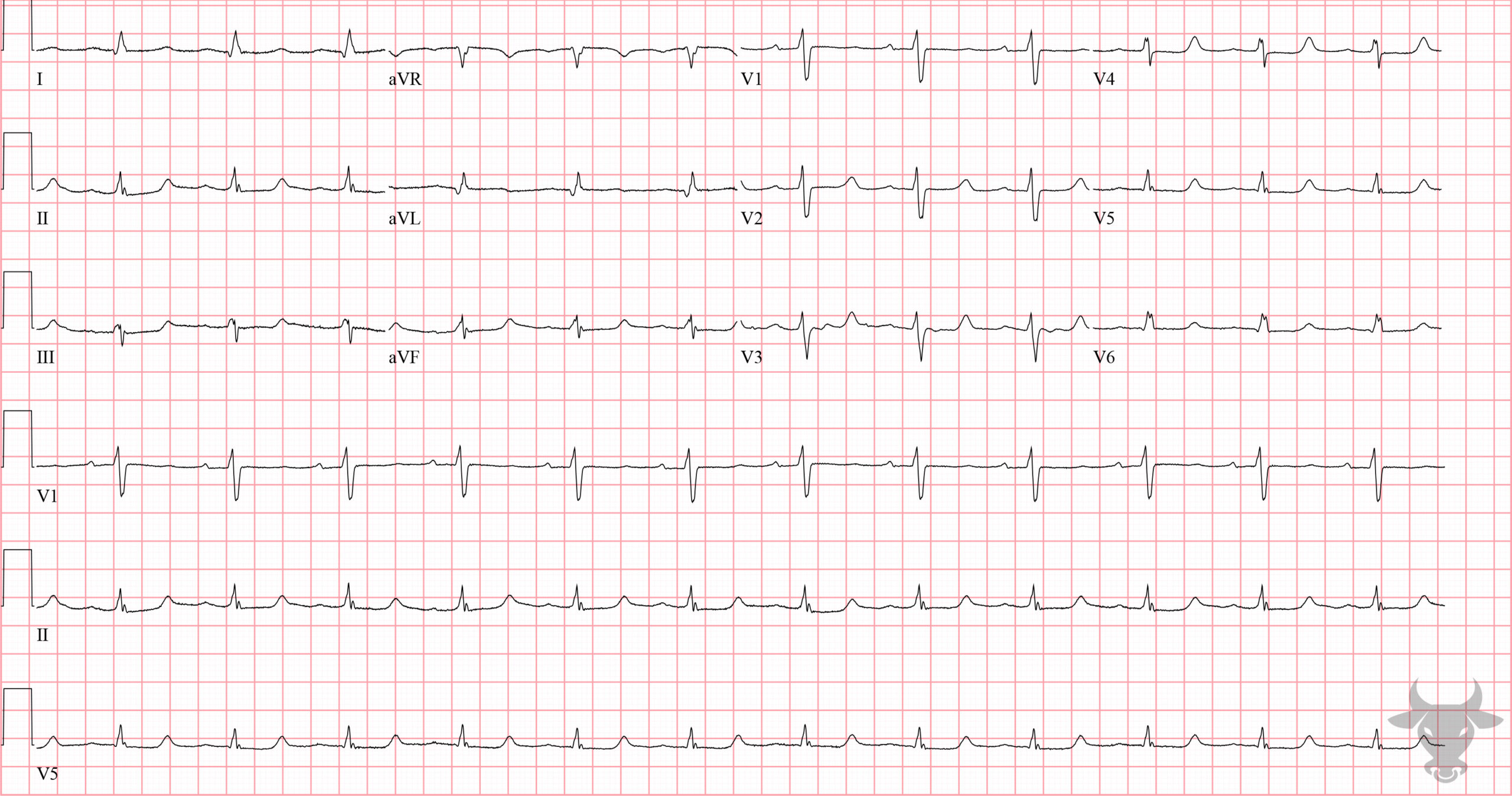
Hypocalcemia
This ECG is classic for end-stage-renal-disease. Notice the narrow-based T waves and lengthened ST segment characteristic of hyperkalemia and hypocalcemia, respectively.
Hypocalcemia
The QT is prolonged by way of a lengthened ST segment. There are narrow-based and a slightly peaked morphology to the T waves (suggesting hyperkalemia). This is a classic appearance of a patient with end-stage-renal-disease.References
- Diercks DB, Shumaik GM, Harrigan RA, Brady WJ, Chan TC. Electrocardiographic manifestations: Electrolyte abnormalities. Journal of Emergency Medicine. 2004;27(2):153-160.