Multilead ST-segment depression with ST-segment elevation in aVR is a pattern that has been recognized as a strong predictor of left main coronary artery or 3-vessel disease; however, occlusive coronary artery disease is not the only cause of this ECG pattern. Frequently, this pattern results from nonocclusive causes such as baseline left ventricular hypertrophy or conditions that create a supply-demand mismatch such as acute blood loss, sepsis, respiratory failure, tachydysrhythmias, and aortic stenosis. In one series of 133 patients showing this ECG pattern, only 28% had acute coronary syndromes, whereas 45% had hypertensive heart disease.
Global Subendocardial Ischemia

Examples
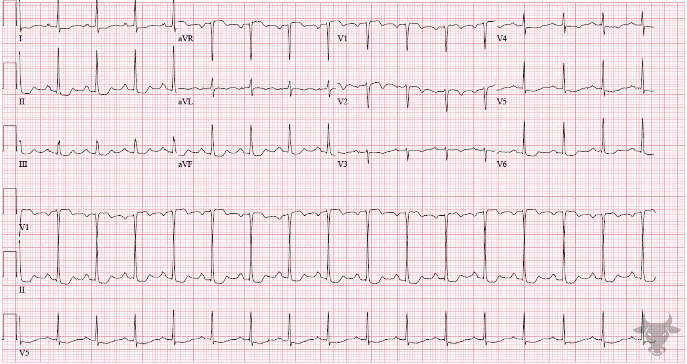
Global Subendocardial Ischemia Due to Multivessel Disease
Multilead ST depressions with ST elevation in aVR. This patient presented with typical chest pain and was discovered to have diffuse disease on left heart catheterization. The patient underwent coronary artery bypass grafting.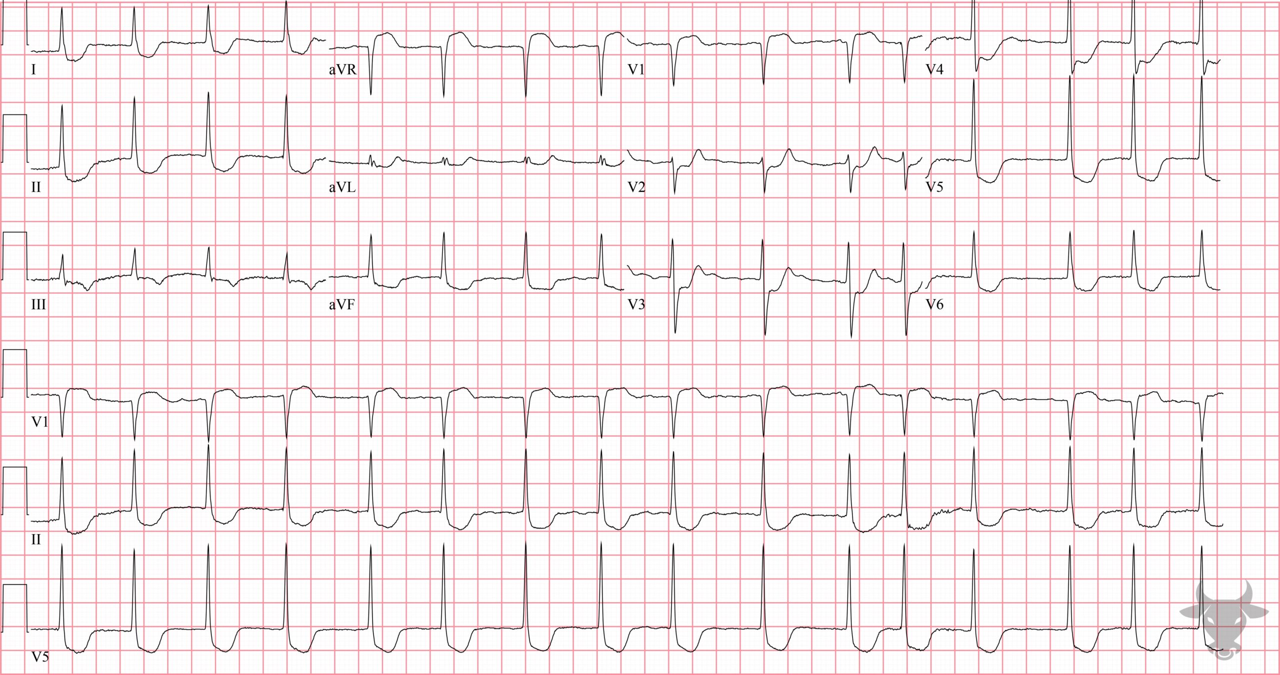
Global Subendocardial Ischemia Due to Multivessel Disease
Multilead ST depressions with ST elevation in aVR. There was diffuse disease discovered on left heart catheterization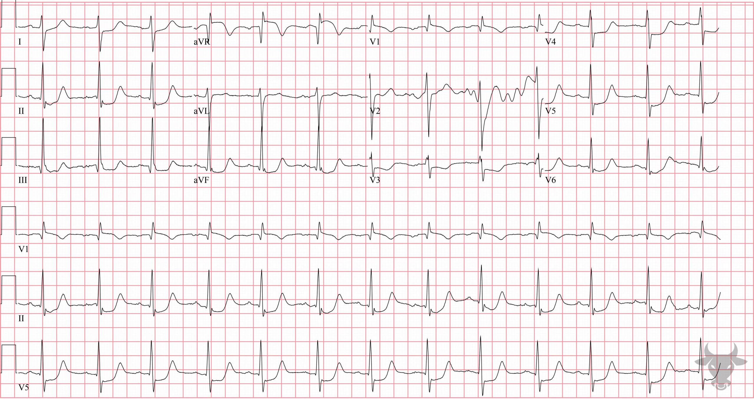
Global Subendocardial Ischemia Due to Cardiac Arrest
Post-return of spontaneous circulation ECG in a cardiac arrest patient. This pattern represents global subendocardial ischemia and can be seen during acute illness or after cardiac arrest.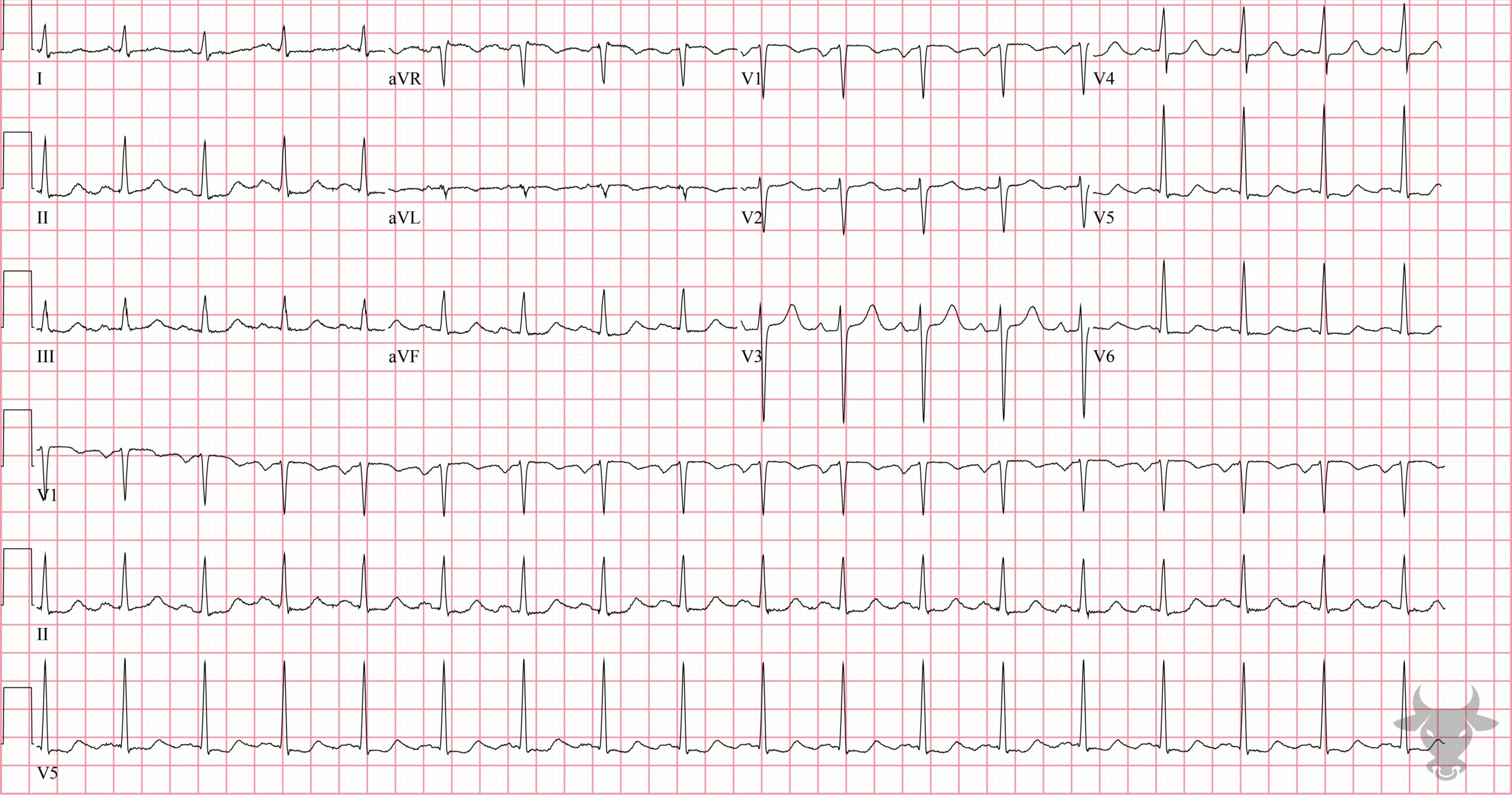
Global Subendocardial Ischemia Due to Severe Anemia
Subtle multilead ST depressions with ST elevation in aVR. This patient had an upper gastrointestinal bleed and was severely anemic, causing this global subendocardial pattern.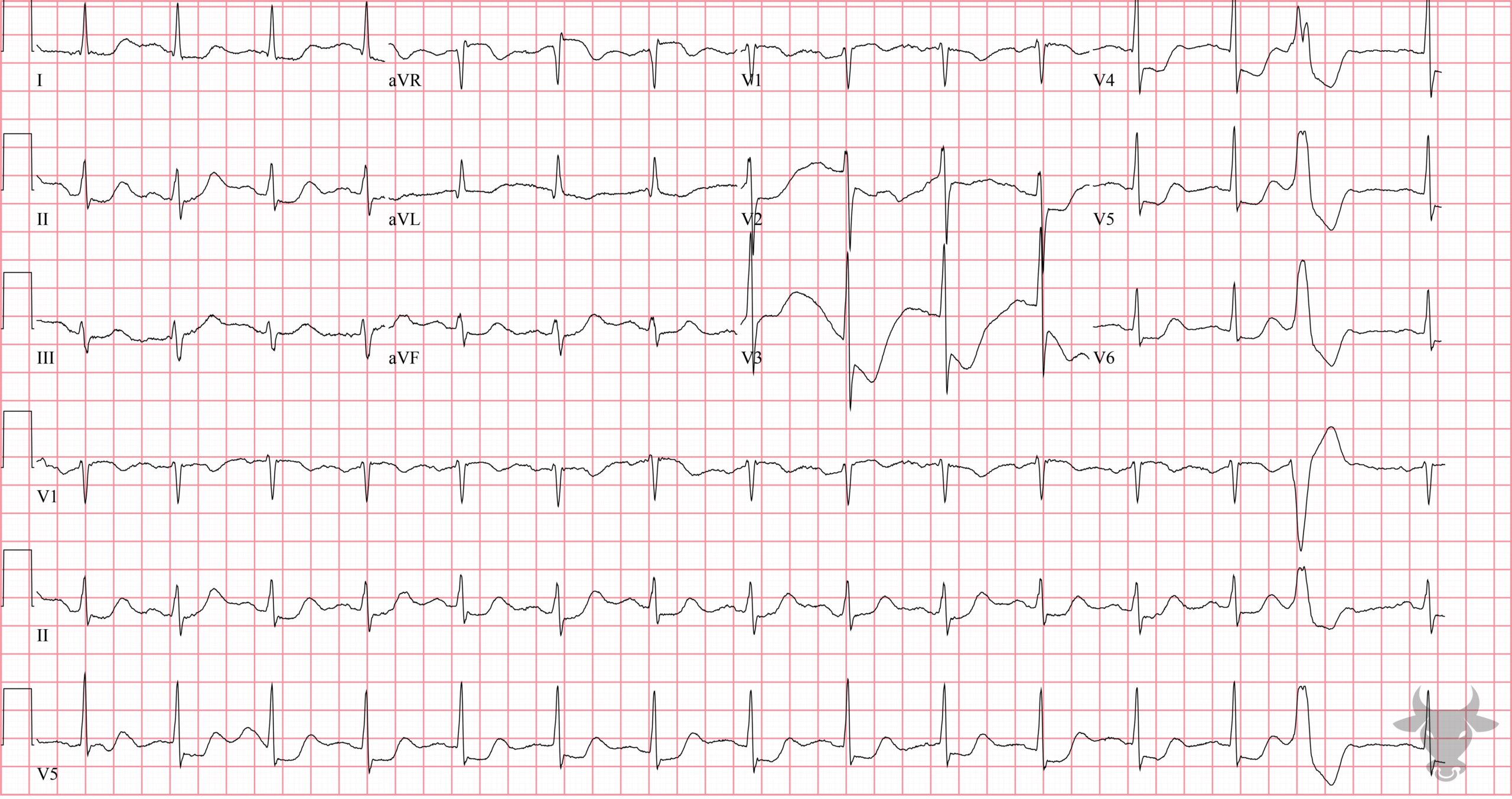
Global Subendocardial Ischemia Due to Aortic Dissection
Multilead ST depression with ST elevation in aVR. This patient was discovered to have a type A aortic dissection.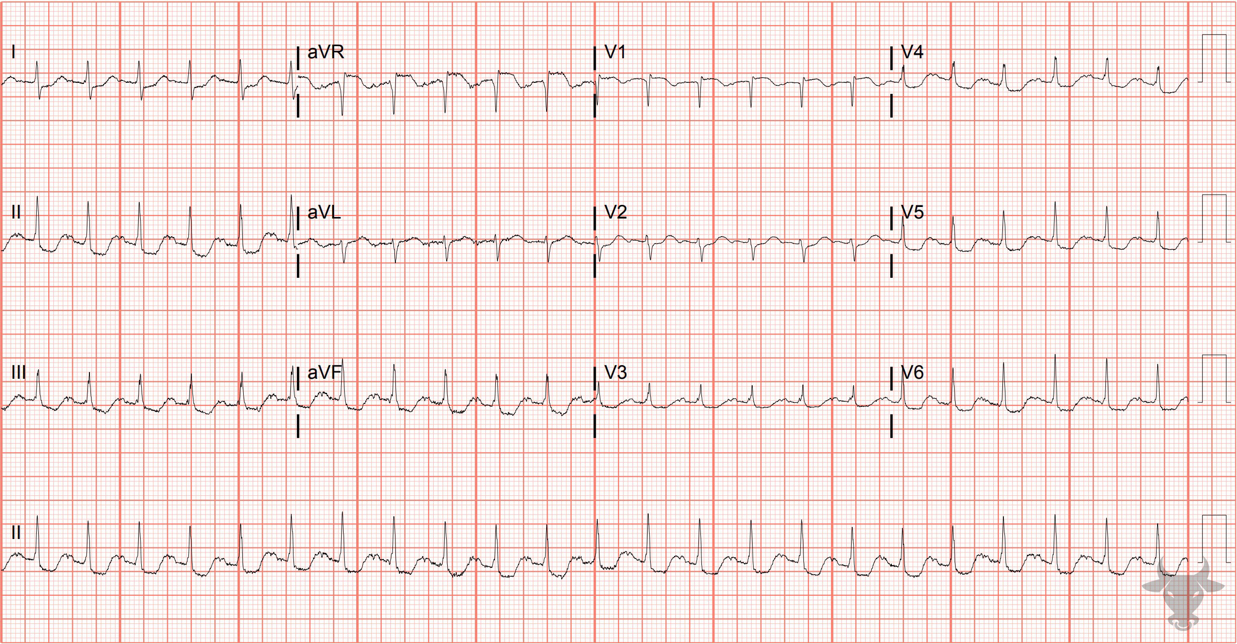
Global Subendocardial Ischemia Due to Pulmonary Embolism
There are diffuse ST-segment depressions along with ST-segment elevation in aVR. This patient was discovered to have a saddle pulmonary embolism for which thrombectomy was performed. The ST changes and tachycardia resolved after mechanical retrieval of the embolus.References
- Witkov RB, Cooper BL. An Ominous ECG Pattern. Annals of Emergency Medicine. 2019;73(4):406-408.
- Miranda D, Lobo A, Walsh B. New insights into the use of the 12-lead electrocardiogram for diagnosing acute myocardial infarction in the emergency department. Canadian Journal of Cardiology. 2018;34:132-145.
- Kukla P, Macintyre W, Fijorek K. Electrocardiographic abnormalities in patients with acute pulmonary embolism complicated by cardiogenic shock. American Journal of Emergency Medicine2. 2014;32:507-510.