Digoxin, or digitalis, is associated with several electrocardiographic changes and, in toxicity, can precipitate a myriad of tachydysrhythmias and bradydysrhythmias. Increased intracellular calcium increases automaticity, and atrioventricular blockade increase vagal tone. This combination can cause atrial tachycardia, junctional rhythms, premature ventricular contractions (most common), sinus bradycardia, and/or slow atrial fibrillation. Bidirectional ventricular tachycardia is near pathognomonic for digoxin toxicity. At therapeutic concentrations it causes scooping of the ST segment predominantly in the precordial leads with tall R waves (V5, V6).
Digoxin Toxicity

Examples
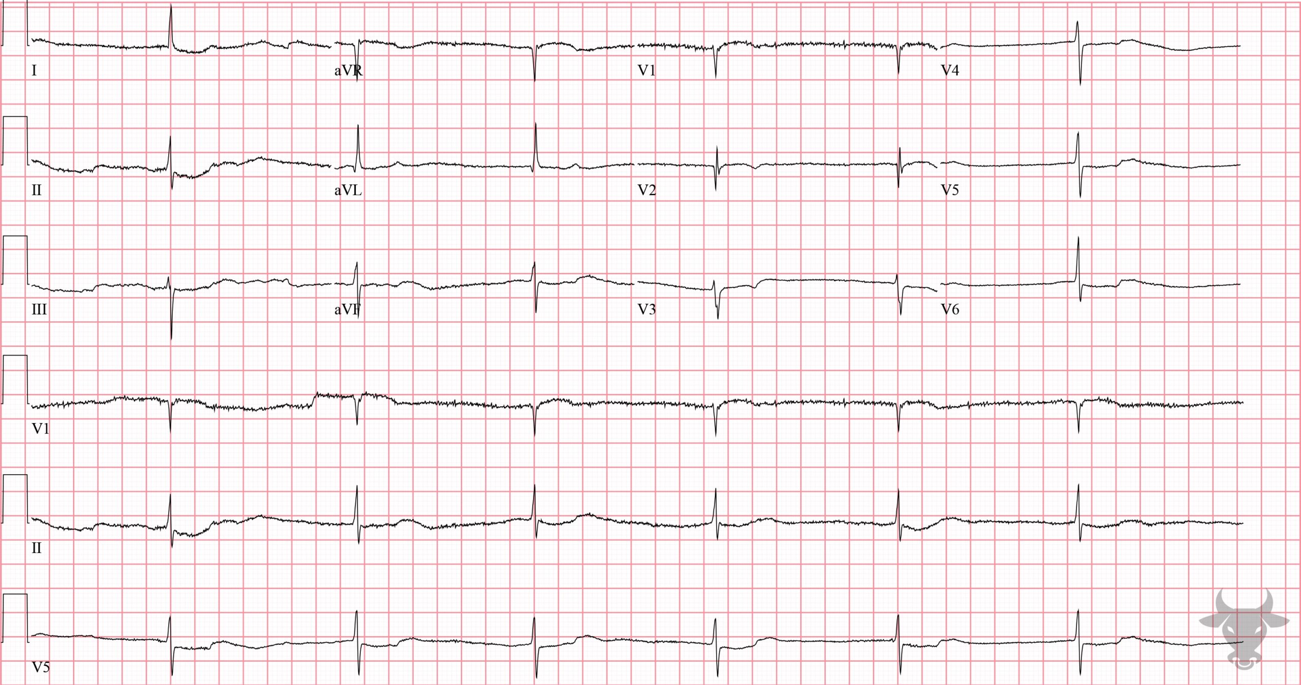
Digoxin Toxicity
This ECG shows the classic "dig effect" (i.e., scooped ST segment) and a junctional bradycardia in a patient with acute renal failure and hyperkalemia.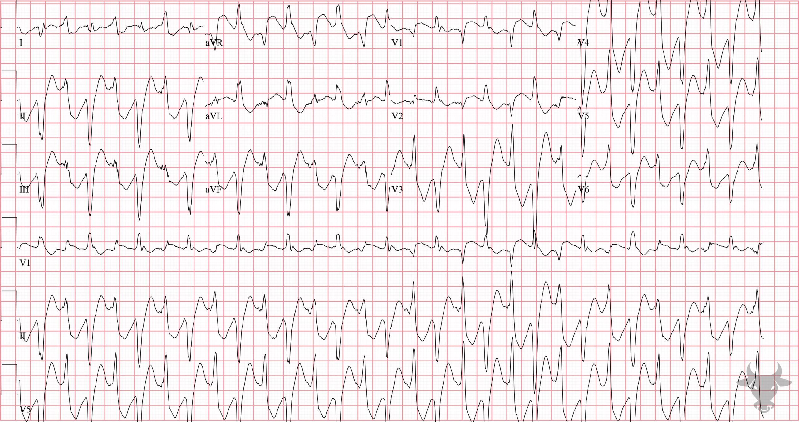
Bidirectional Ventricular Tachycardia
Bidirectional ventricular tachycardia is a type of polymorphic ventricular tachycardia and is near pathognomonic for digoxin toxicity.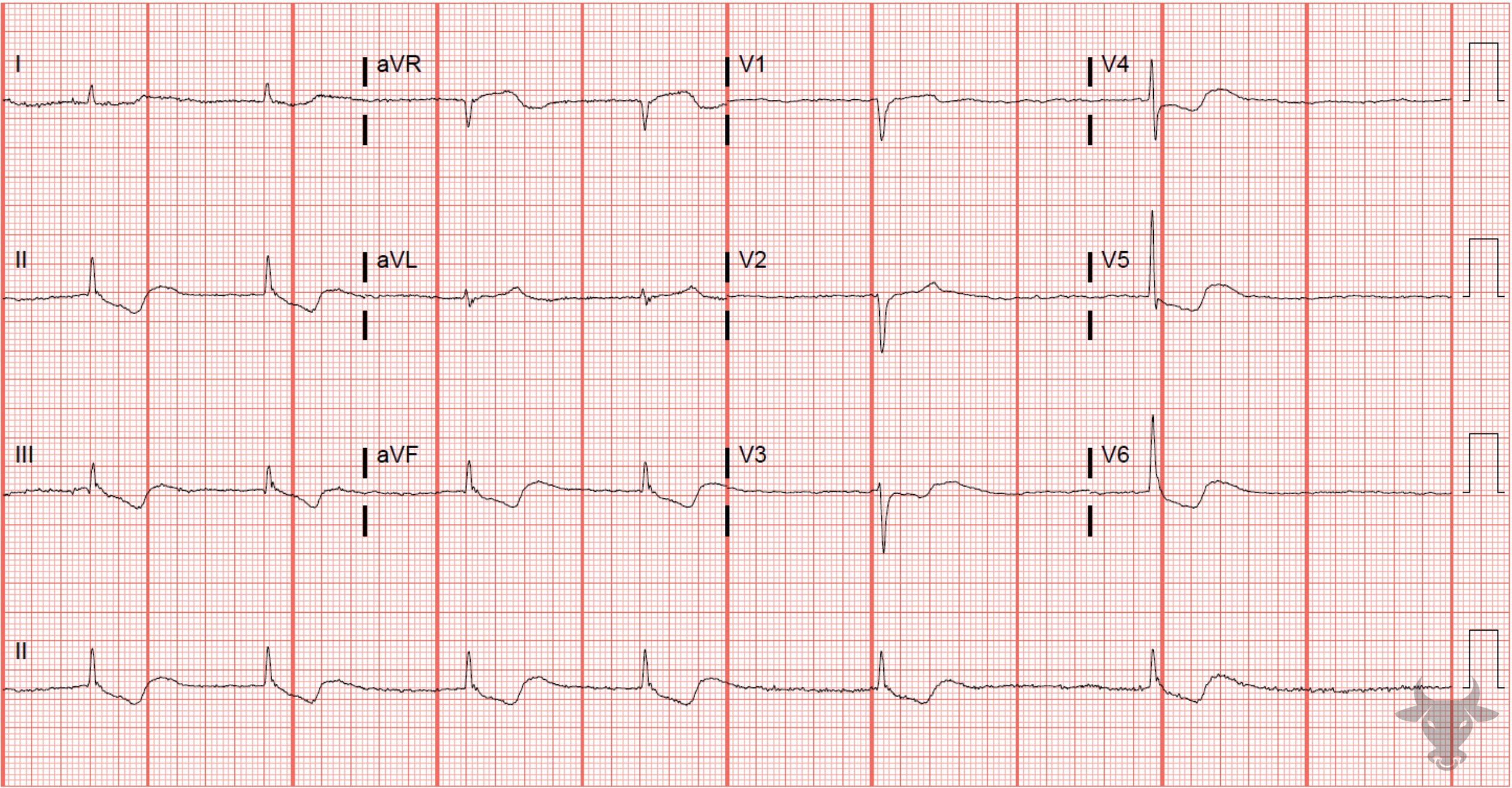
Atrial Fibrillation
Slow atrial fibrillation due to digoxin toxicity. Digoxin slows conduction through the atrioventricular node and increases automaticity. Slow atrial fibrillation is a common rhythm with digoxin toxicity. Also note the "scooped" ST-segments typical of the digoxin-effect.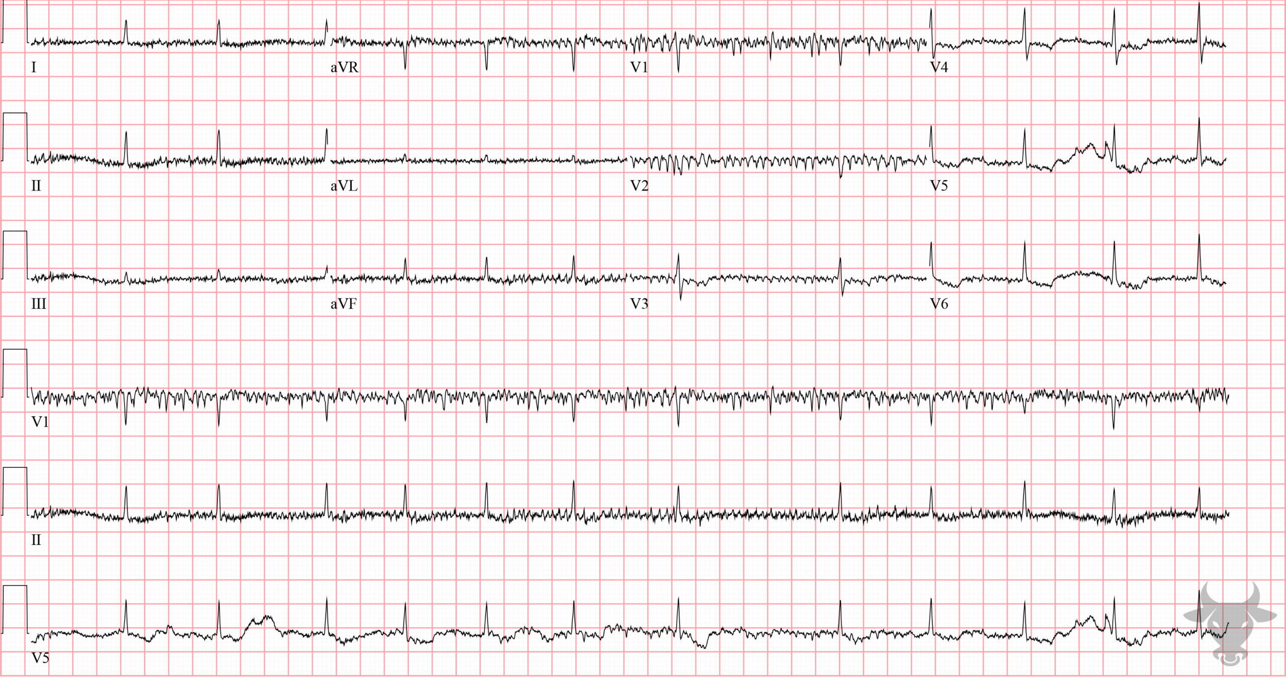
Digoxin Effect
There is atrial fibrillation with baseline artifact, and a sagging or scooped ST segment best seen in the lateral precordial leads. These findings are expected of someone who takes digoxin, which this patient did.References
1. Mattu A, Tabas J, Brady W. Electrocardiography in Emergency, Acute, and Critical Care. 2nd ed. Dallas, TX: The American College of Emergency Physicians; 2019.