Atrioventricular nodal reentrant tachycardia (AVNRT) is caused by a reentrant loop within the atrioventricular node. With AVNRT, the atrioventricular node has two pathways, fast and slow, which allows for a reentrant loop. The differential for a regular, narrow complex tachycardia includes sinus tachycardia, atrioventricular nodal reentrant tachycardia (AVNRT), atrioventricular reentrant tachycardia (AVRT), atrial flutter, and atrial tachycardia. P waves can aid the diagnosis but are often absent. At faster rates, sinus tachycardia can be obscured when P waves are buried within the T waves. P waves in a sawtooth pattern favors atrial flutter (2:1 conduction usually has a ventricular response rate around 150 bpm). While most cases of AVNRT do not have visible P waves, up to one third of AVNRT cases will show retrograde P’ waves immediately following the QRS complex, giving the appearance of a “pseudo-S wave” in the inferior limb leads, or a “pseudo-R wave” in V1. Rarely, atypical “fast-slow” AVNRT can produce retrograde P’ waves that precede the QRS complex.
Atrioventricular Nodal Reentrant Tachycardia

Examples
Atrioventricular Nodal Reentrant Tachycardia
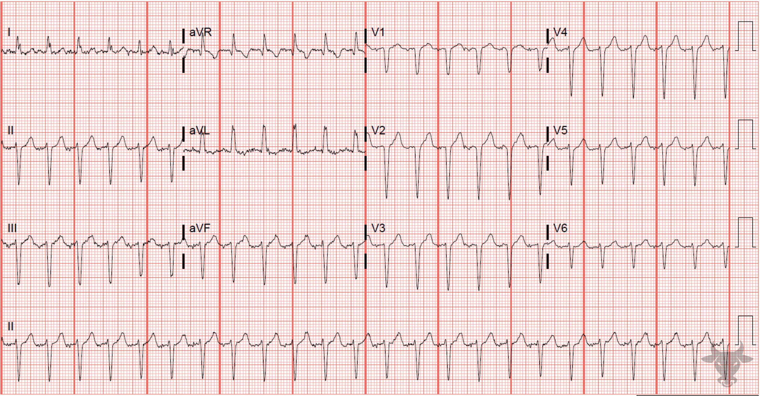
Typical atrioventricular nodal reentrant tachycardia conducts down the slow pathway and up the fast pathway all within the node, and retrograde P waves can occasionally be seen immediately following the QRS complex. In atypical atrioventricular tachycardia, which conducts down the fast and up the slow pathway, retrograde atrial activity can occasionally be seen prior to or in-between QRS complexes as with this case. This patient converted with adenosine helping to confirm the diagnosis of atrioventricular tachycardia.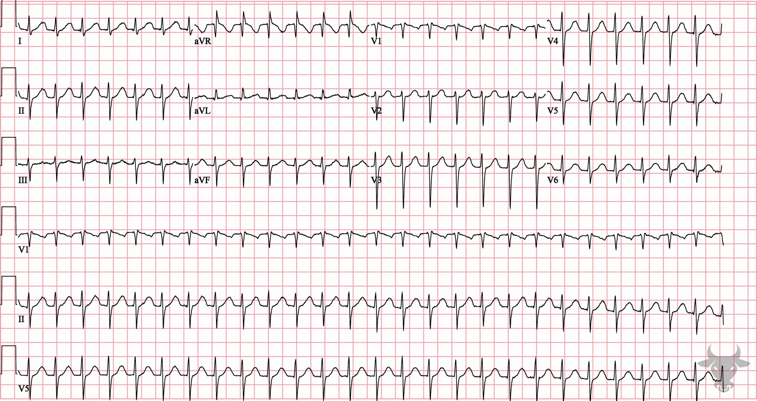
Atrioventricular Nodal Reentrant Tachycardia
Atrioventricular nodal reentrant tachycardia that converted with adenosine.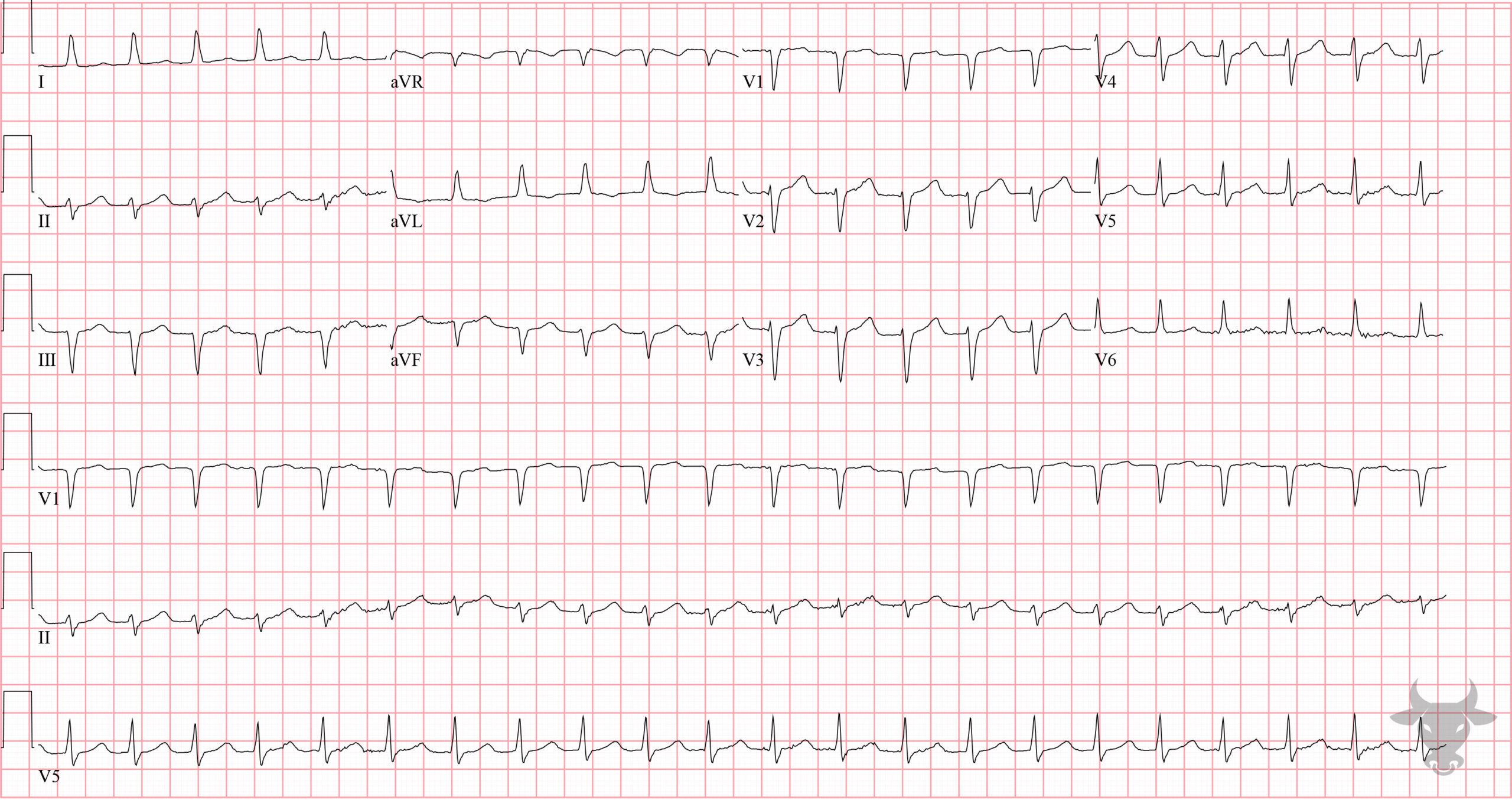
Atrioventricular Nodal Reentrant Tachycardia
This patient converted to sinus rhythm with 12 mg of adenosine. Most textbooks report that the lower rate limit of supraventricular tachycardia is 140 to 150 bpm, but this is not anecdotally true as this case demonstrates.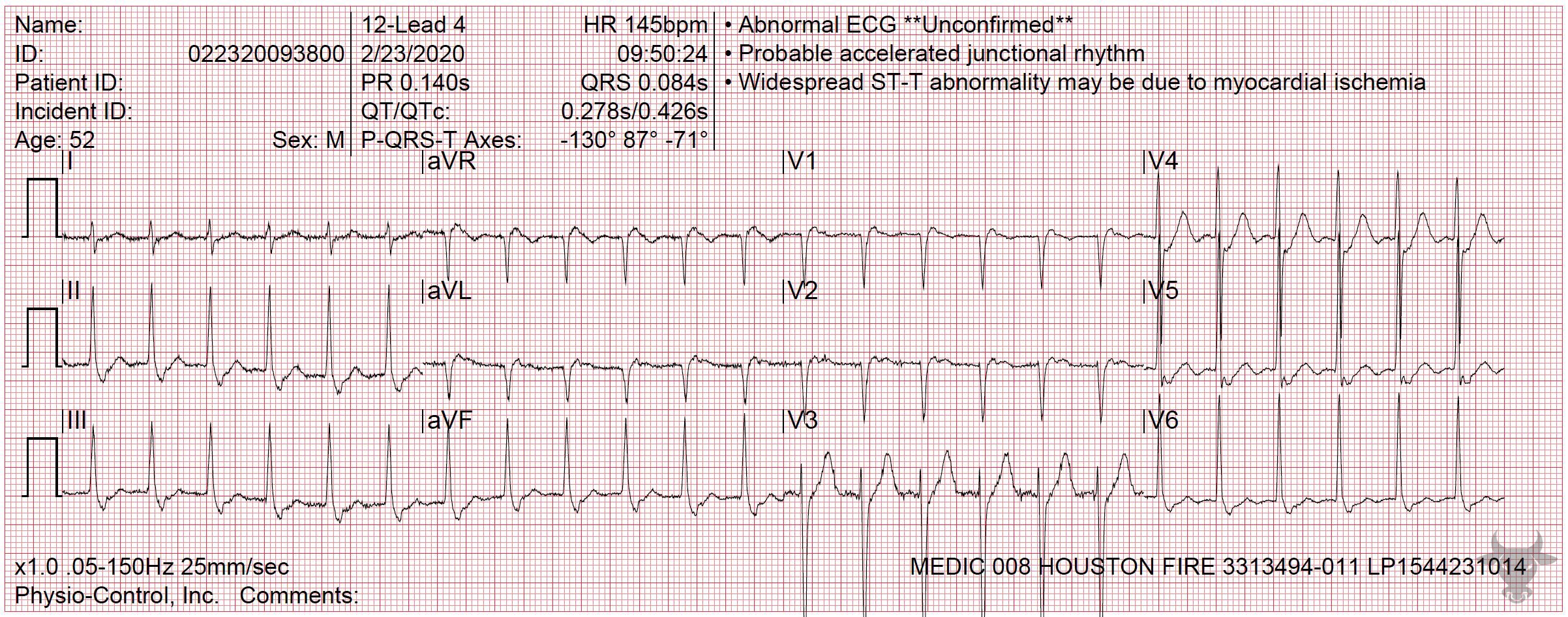
Atrioventricular Nodal Reentrant Tachycardia
Retrograde P waves are seen immediately following the QRS complexes in leads V1 and II, and appear like pseudo-R and pseudo-S waves, respectively.References
- Wagner GS, Strauss DG. Marriott’s Practical Electrocardiography. 12th ed. Lippincott Williams & Wilkins; 2014
- Katritsis DG, Camm AJ. Atrioventricular nodal reentrant tachycardia. Circulation. 2010;122(8):831-840.
- Cooper BL, Beyene JA. Atrioventricular nodal reentrant tachycardia and cannon A waves: A case report. American Journal of Emergency Medicine. Published online 2018.