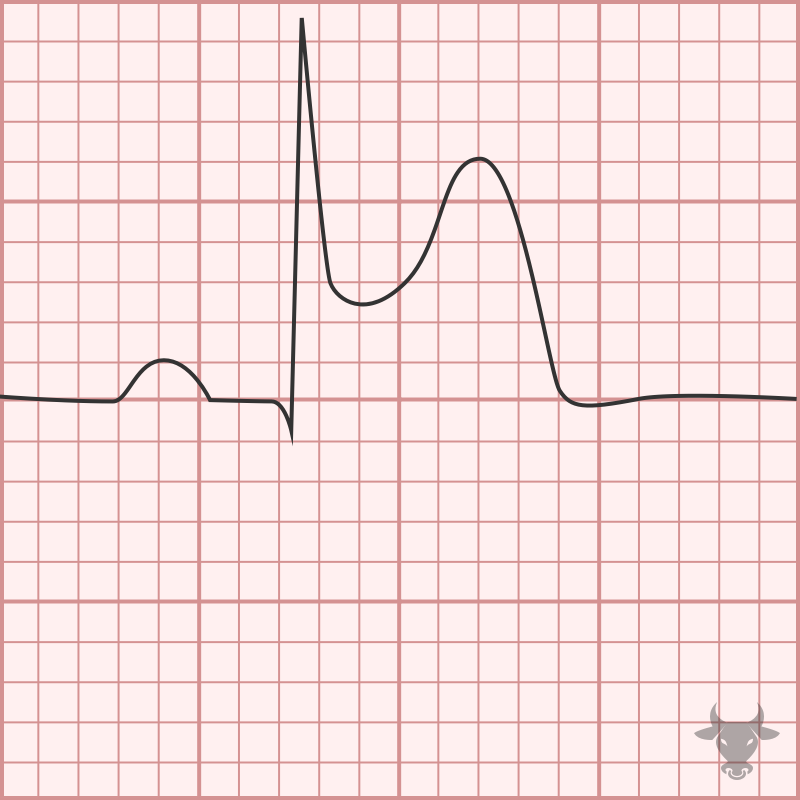
Anterior ST-elevation Myocardial Infarction
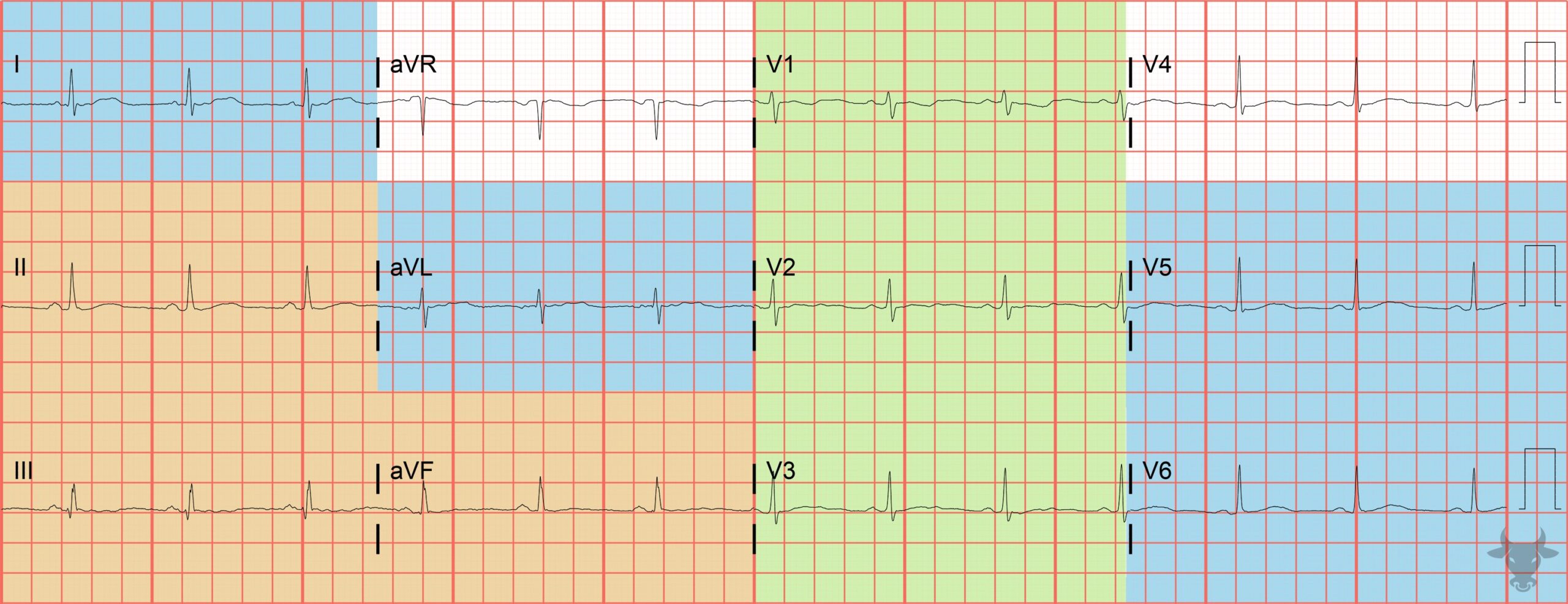
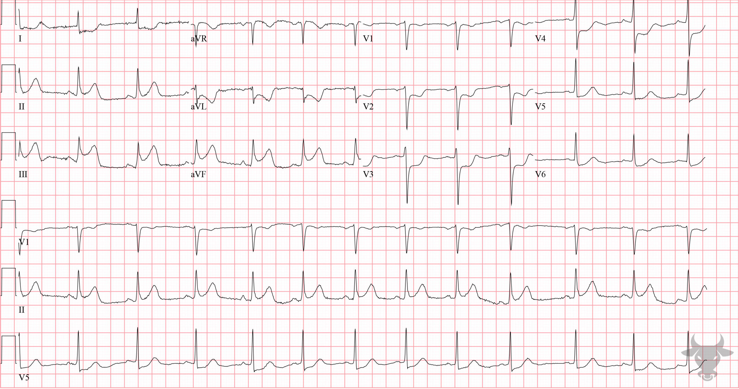
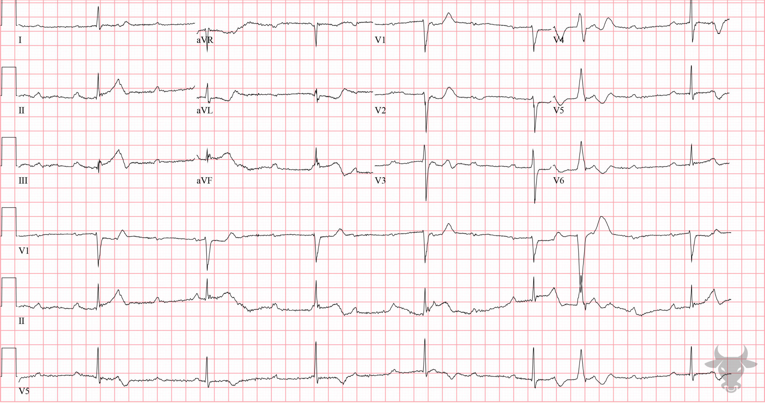
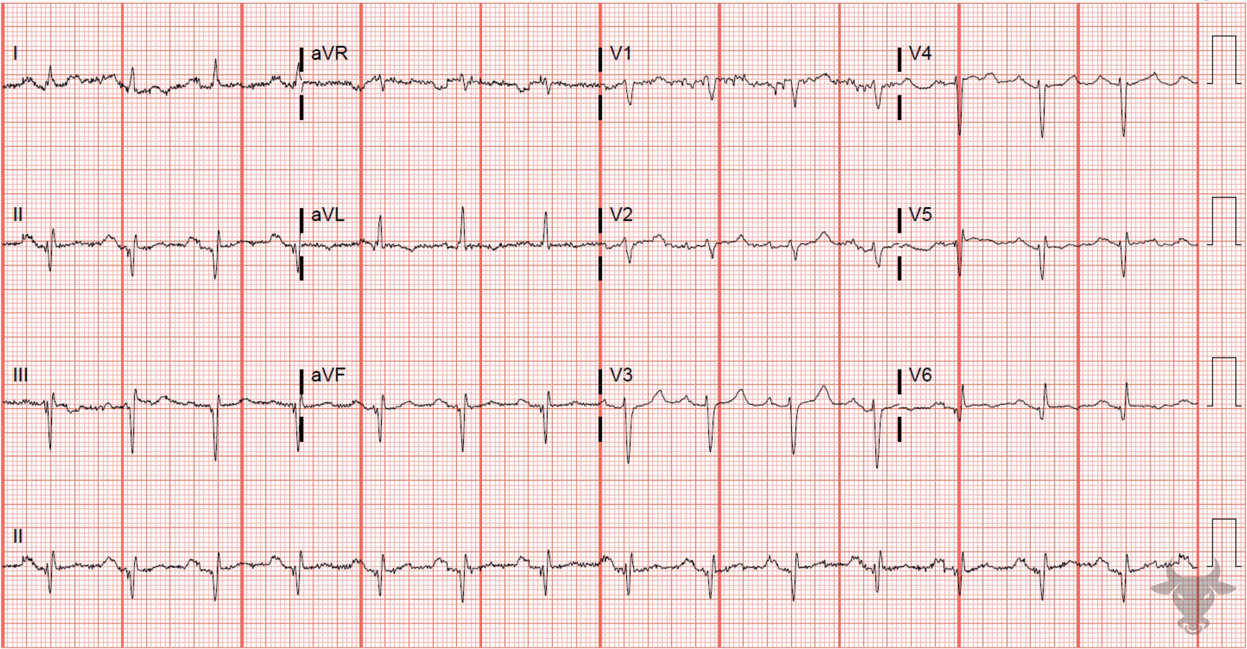
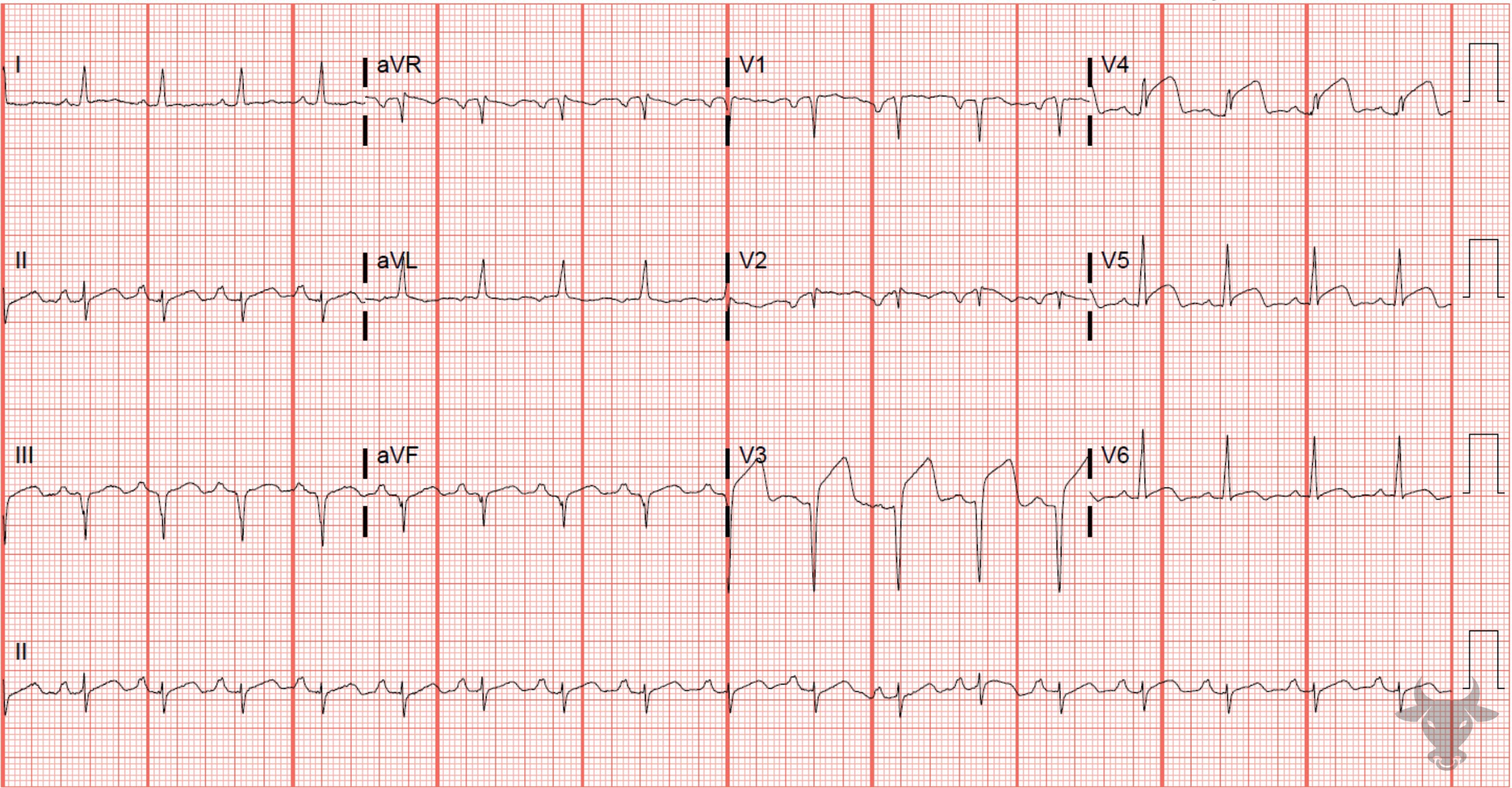
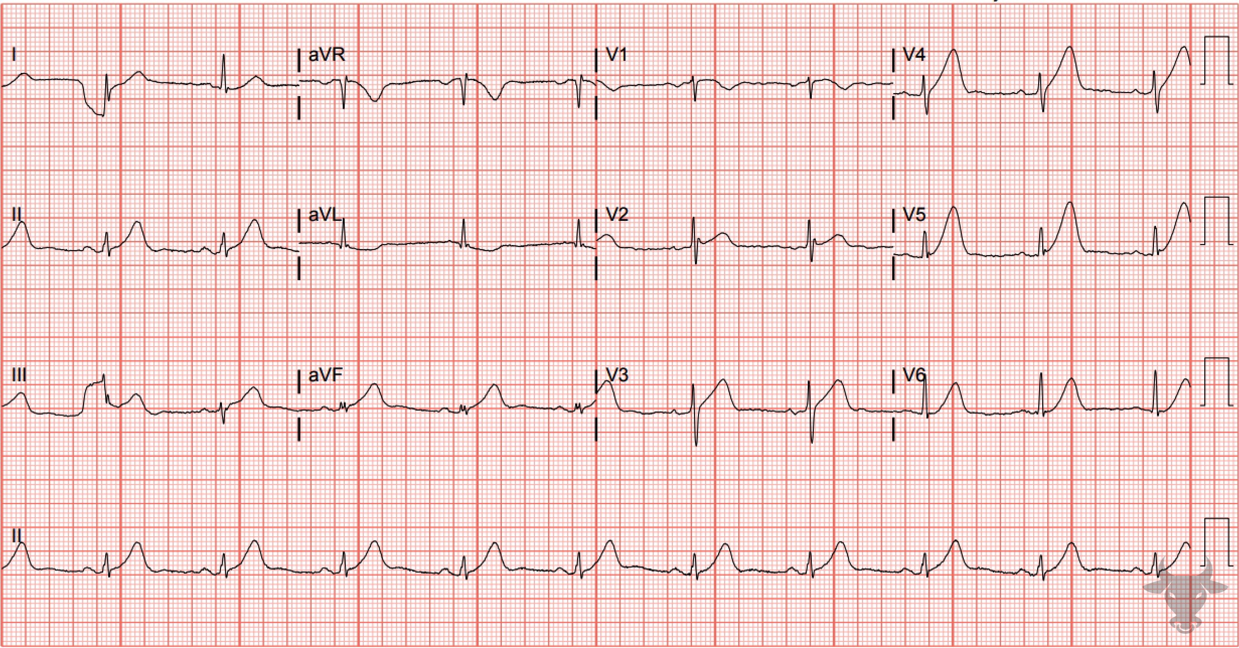
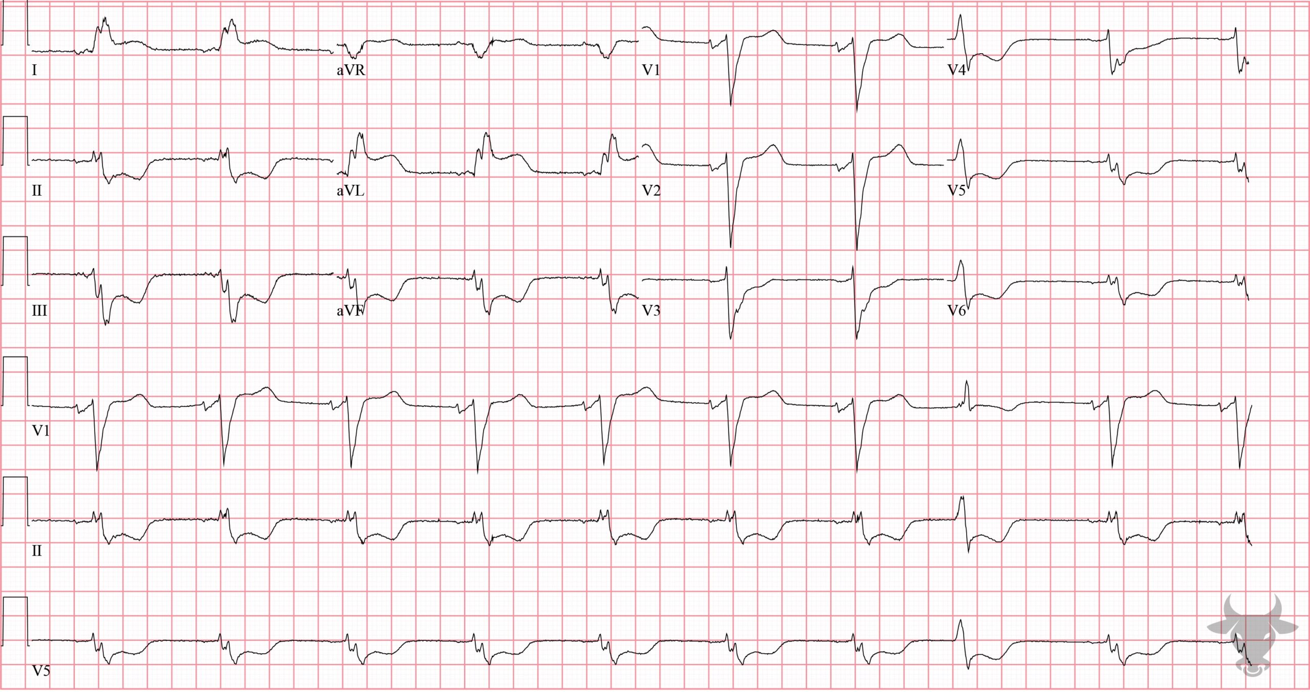
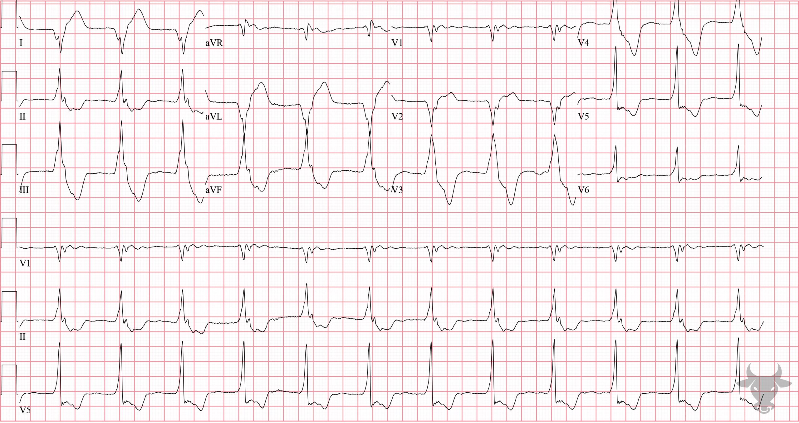
Pay attention to the natively conducted complexes when interpreting this ECG (i.e., ignore the premature ventricular complexes when looking for acutely ischemic findings). There are anterior ST-segment elevations (V1 through V4) and reciprocal ST-segment depressions in the inferior leads (II, III, and aVF). Left heart catheterization revealed left anterior descending artery occlusion.Anterior ST-elevation Myocardial Infarction
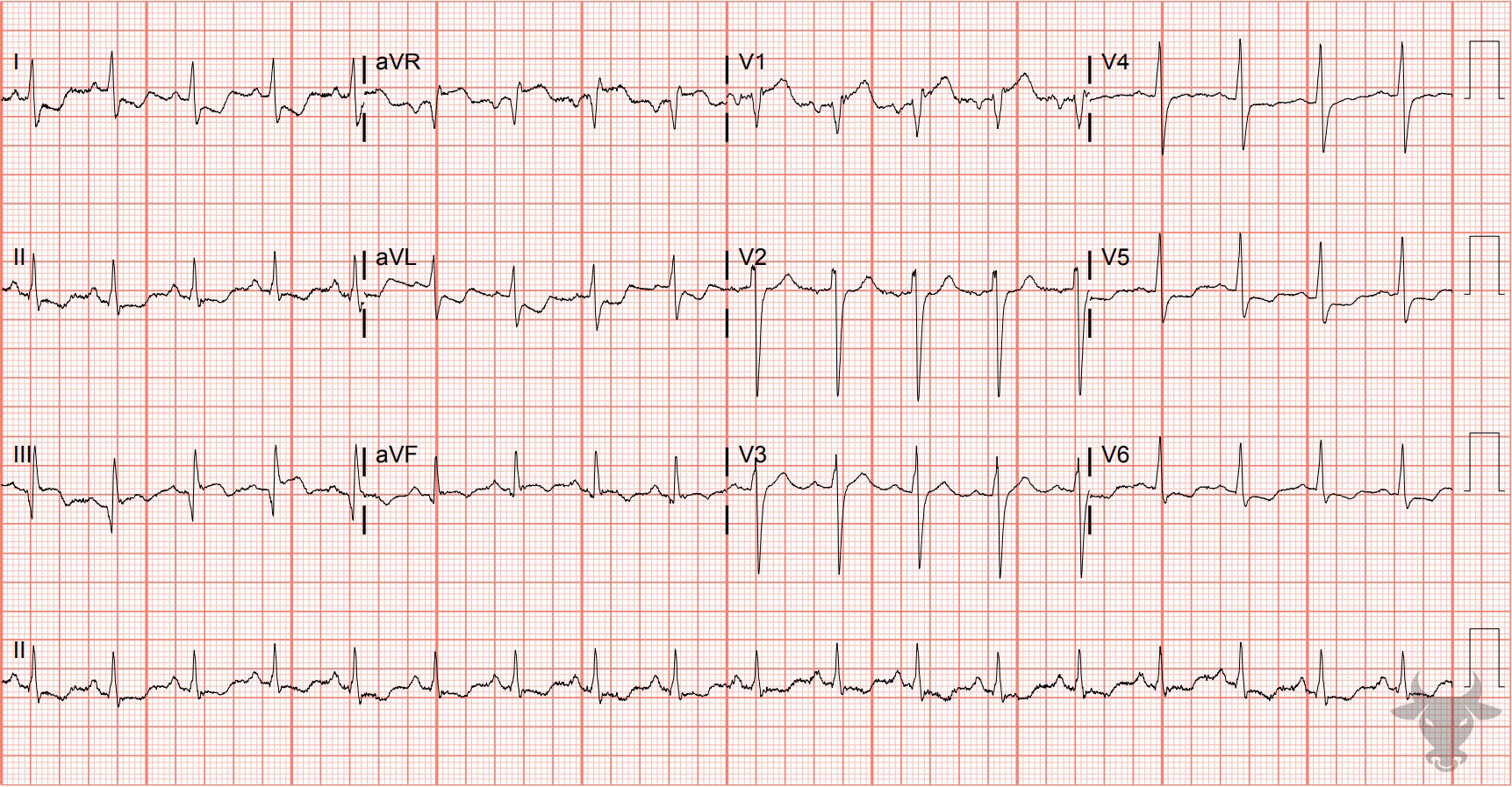
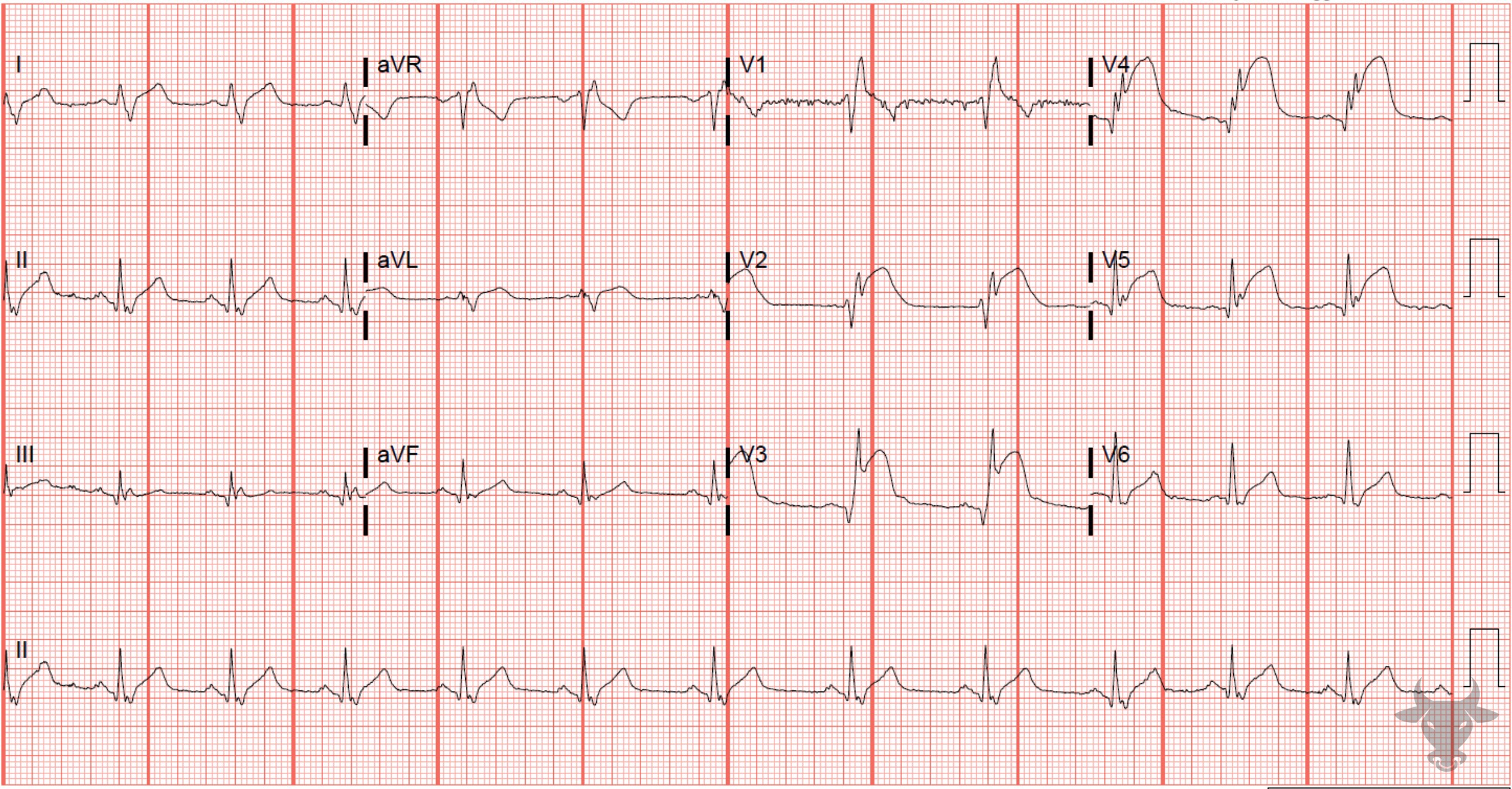
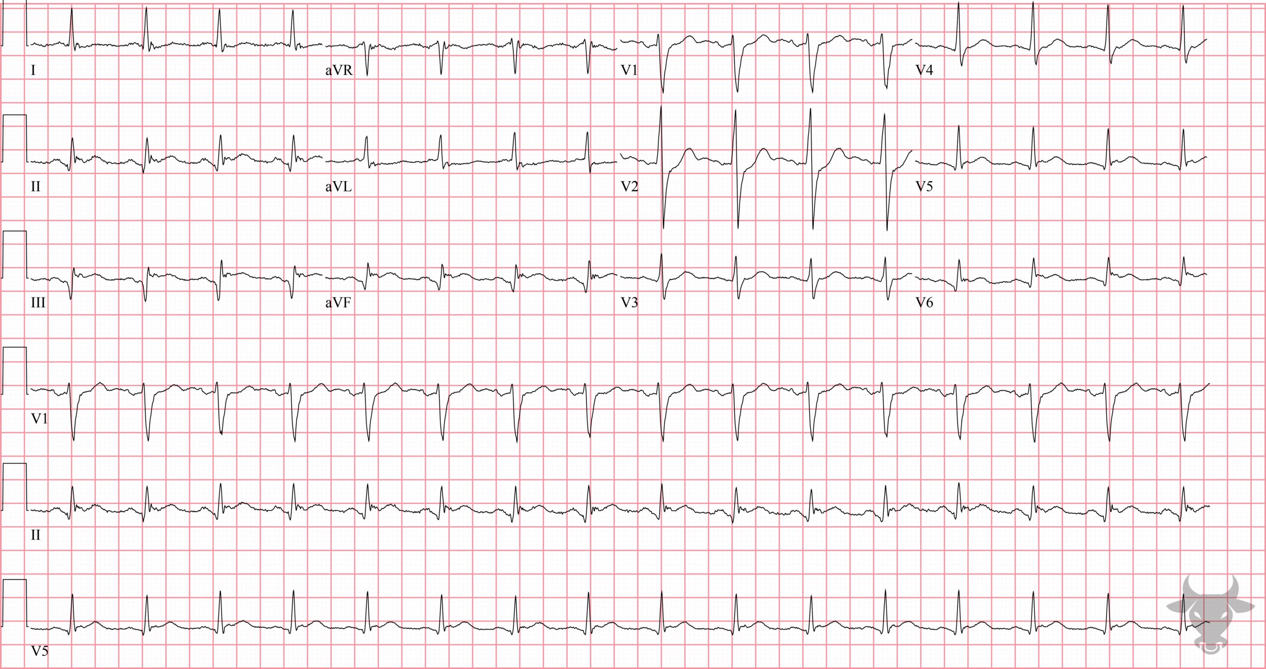
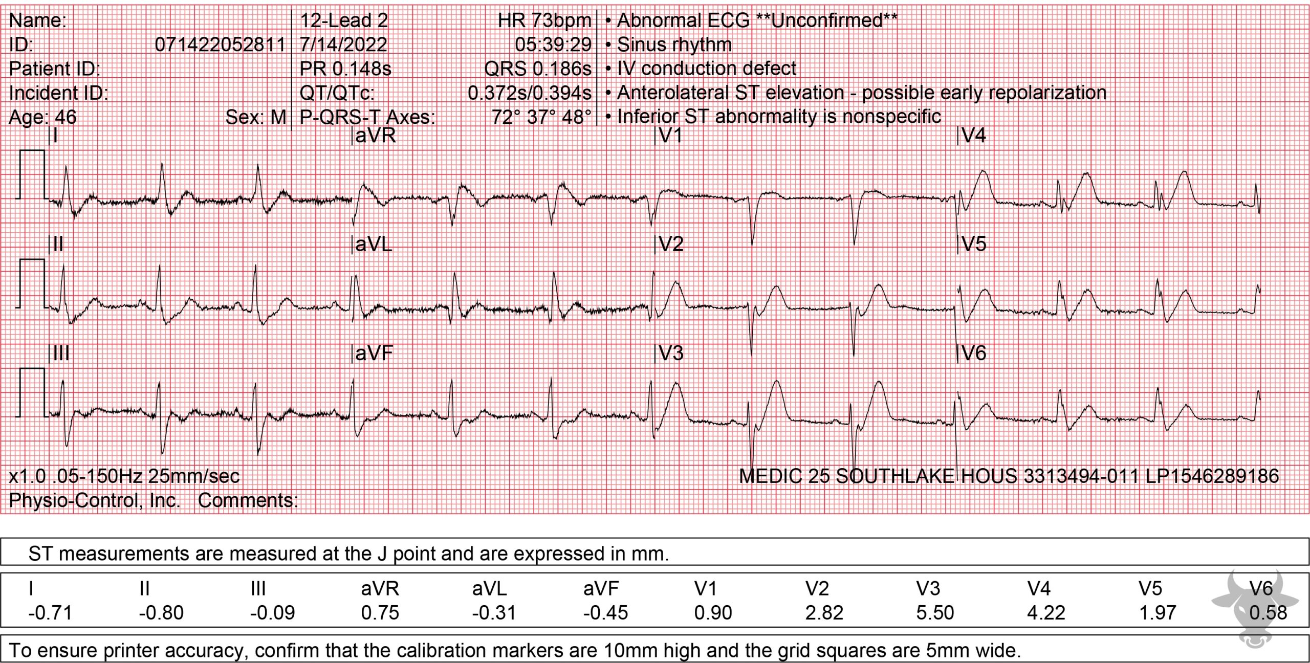
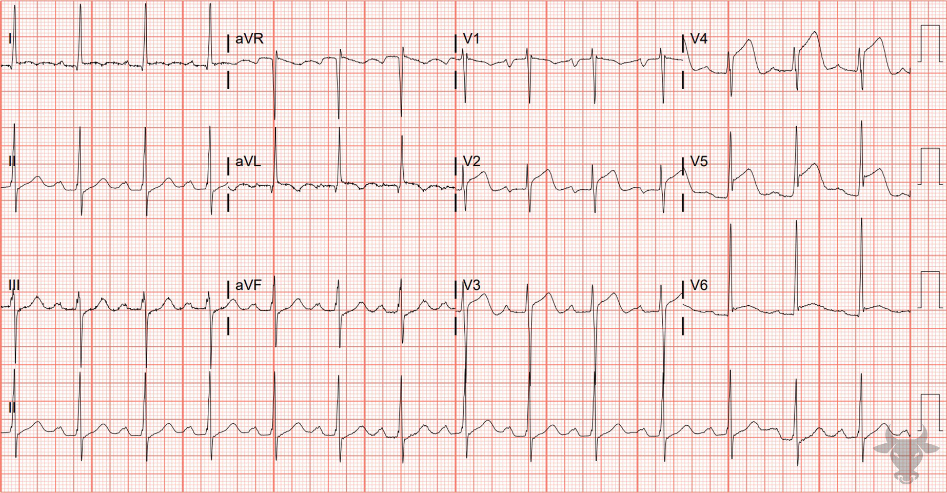
Some STEMIs can be subtle like this one. Notice the slight reciprocal ST depressions in the high lateral leads (i.e., I and aVL). Also note terminal QRS distortion in V3 - the absence of an S wave or a J notch (to suggest early repolarization) - a very specific finding for STEMI. This patient had a 90% mid-left anterior descending artery culprit lesion that was stented.Inferior ST-elevation Myocardial Infarction
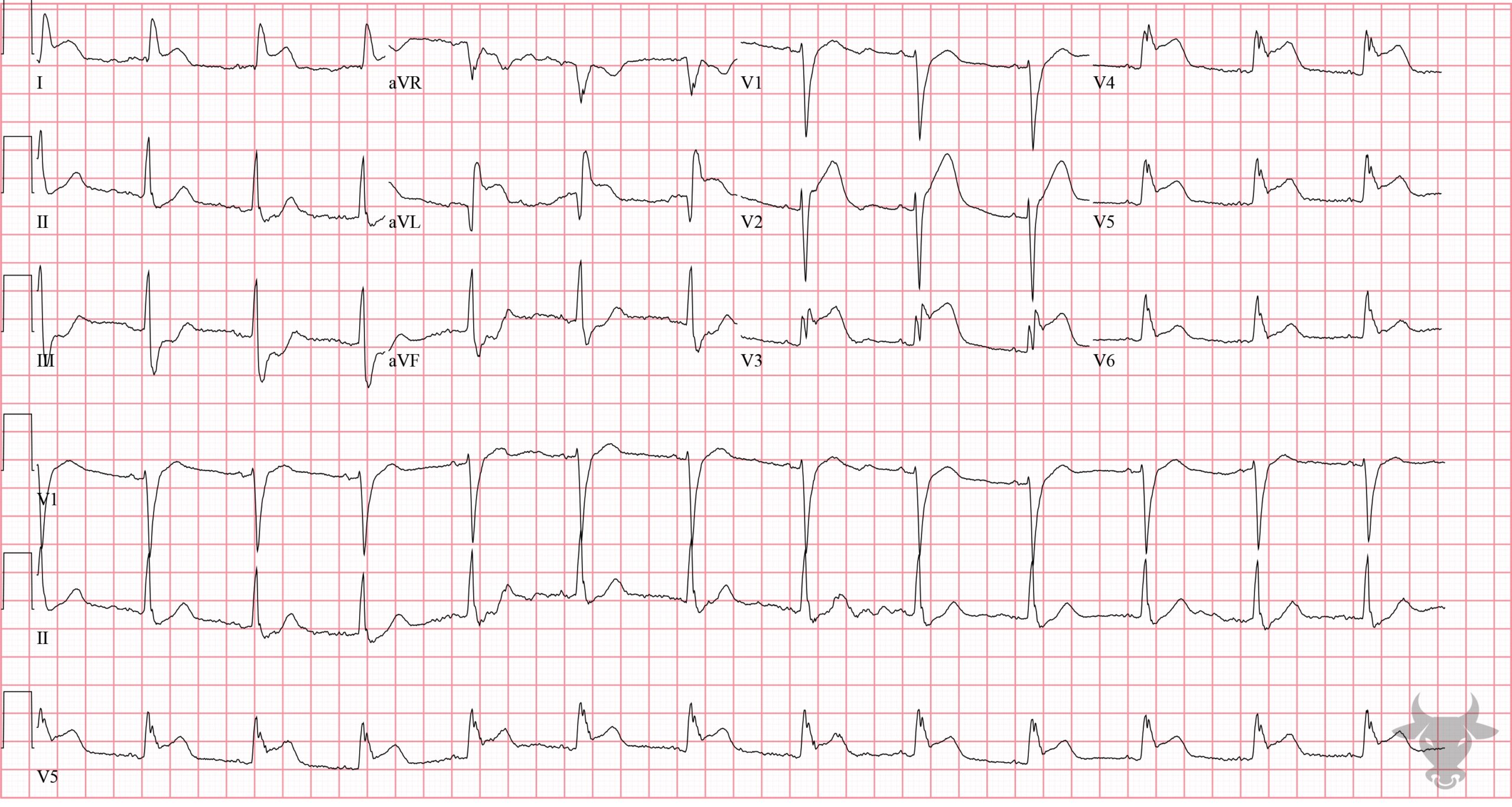
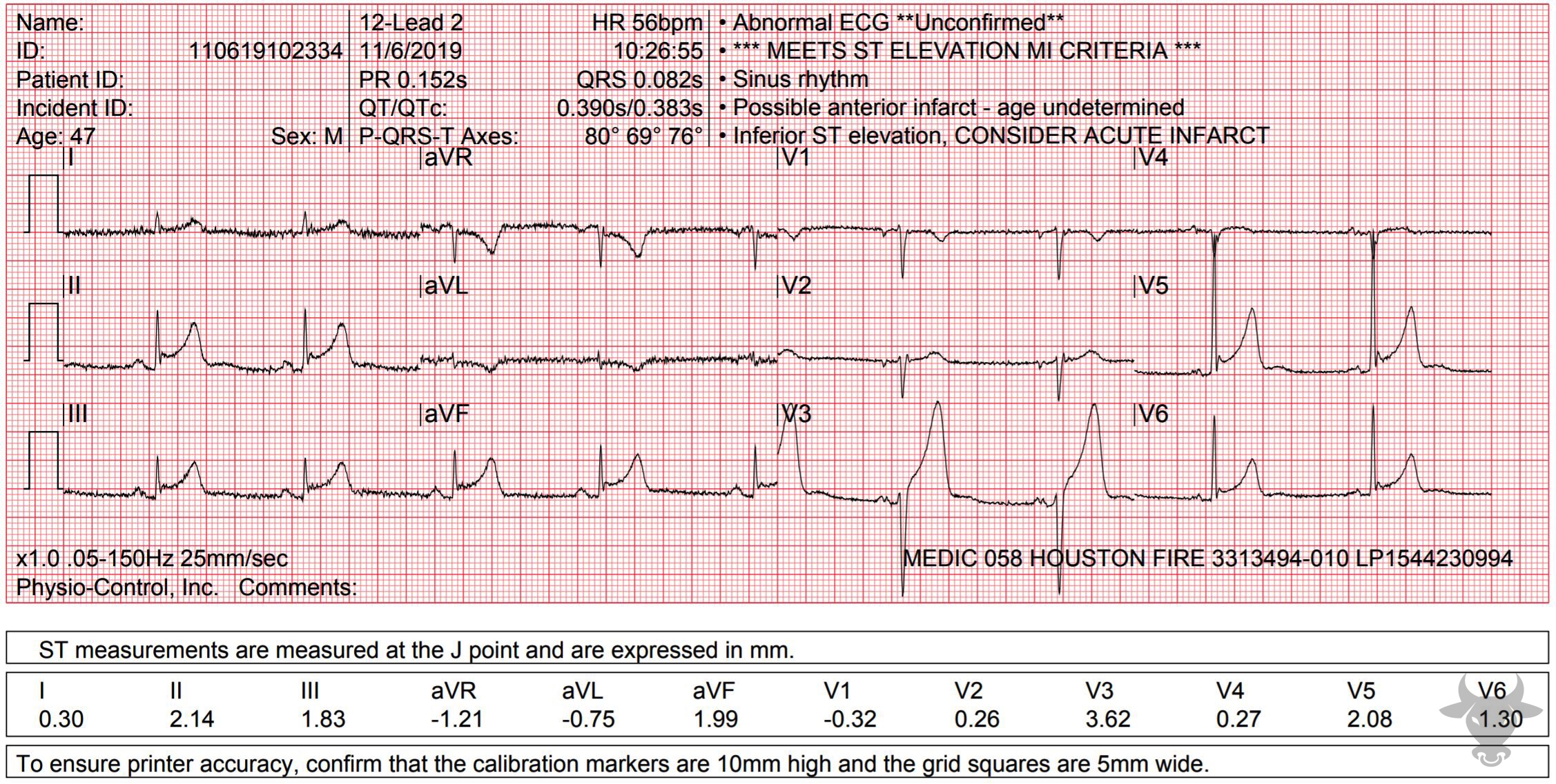
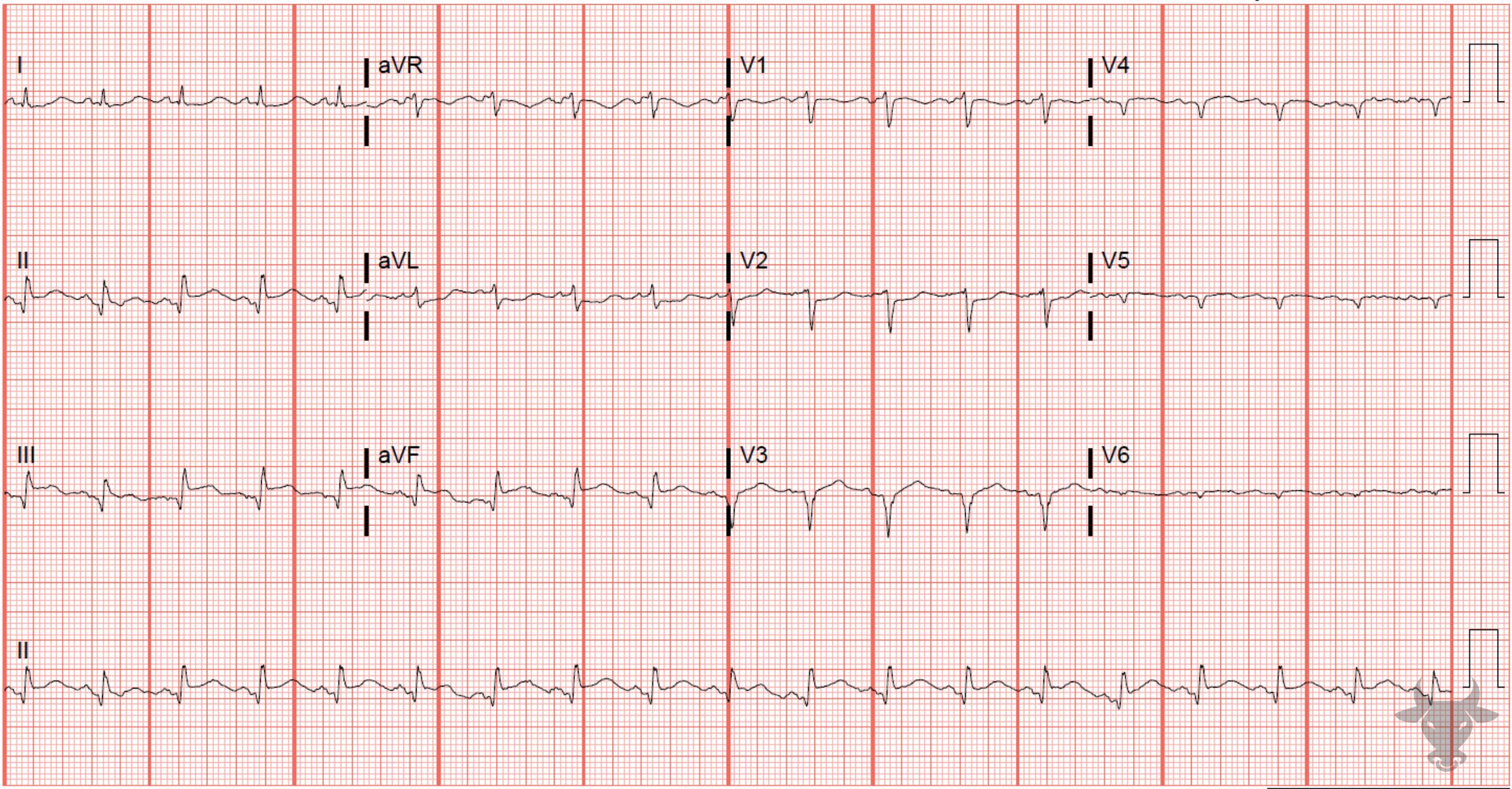
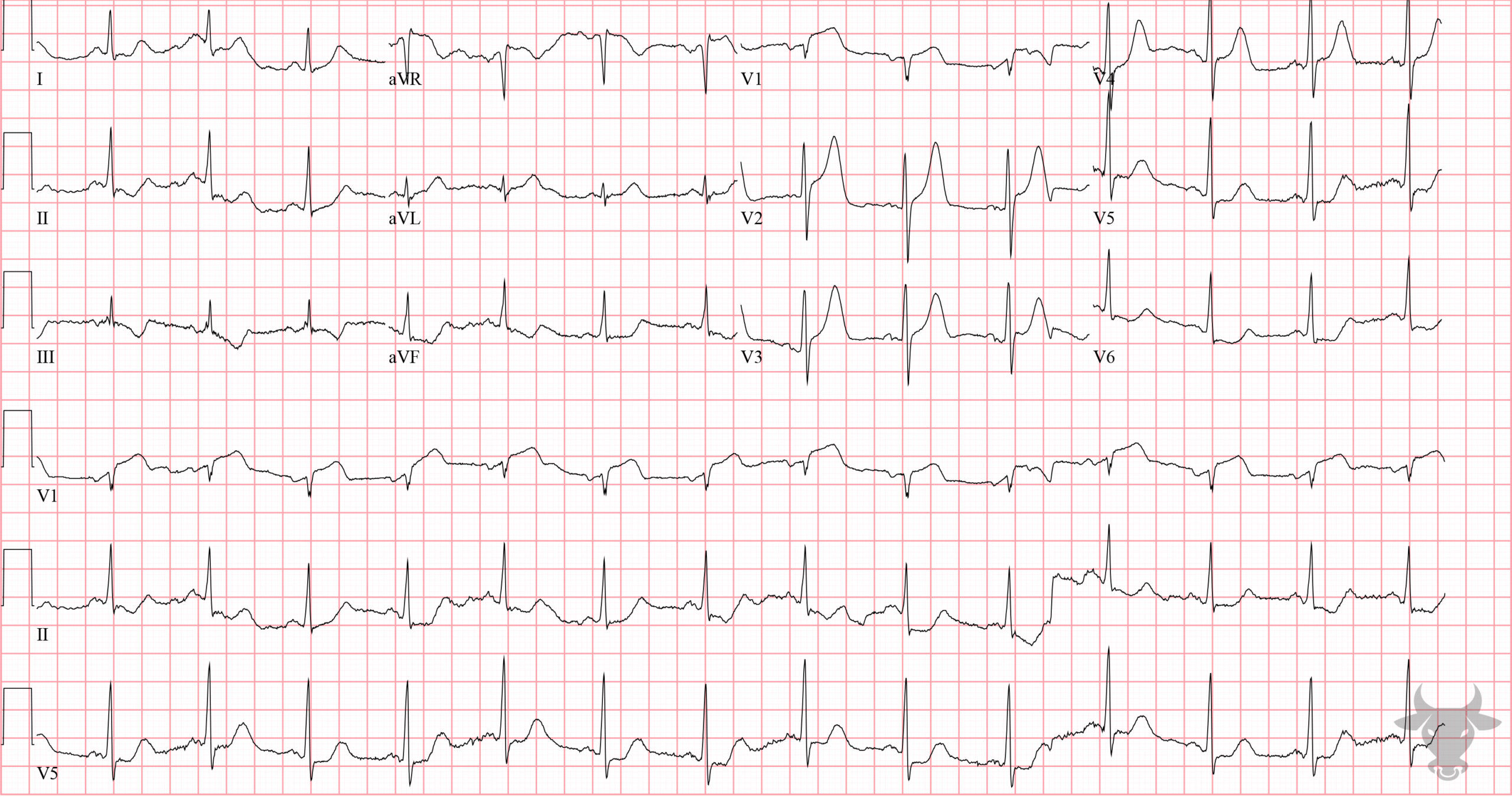
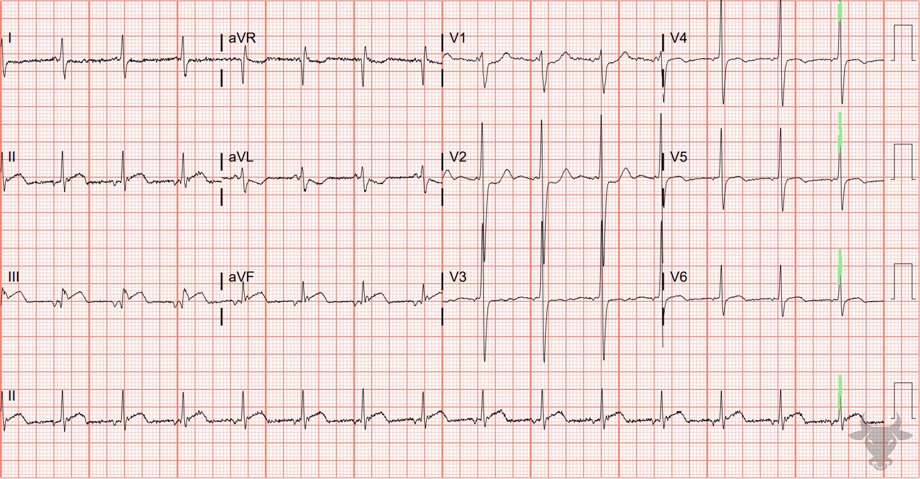
ST-elevation in the inferior leads (II, III, aVF) with reciprocal changes in the anterolateral leads (I, aVL, V2 through V6). This patient received percutaneous coronary intervention to an occluded right coronary artery. ST-elevation Myocardial Infarction (Aslanger Pattern)
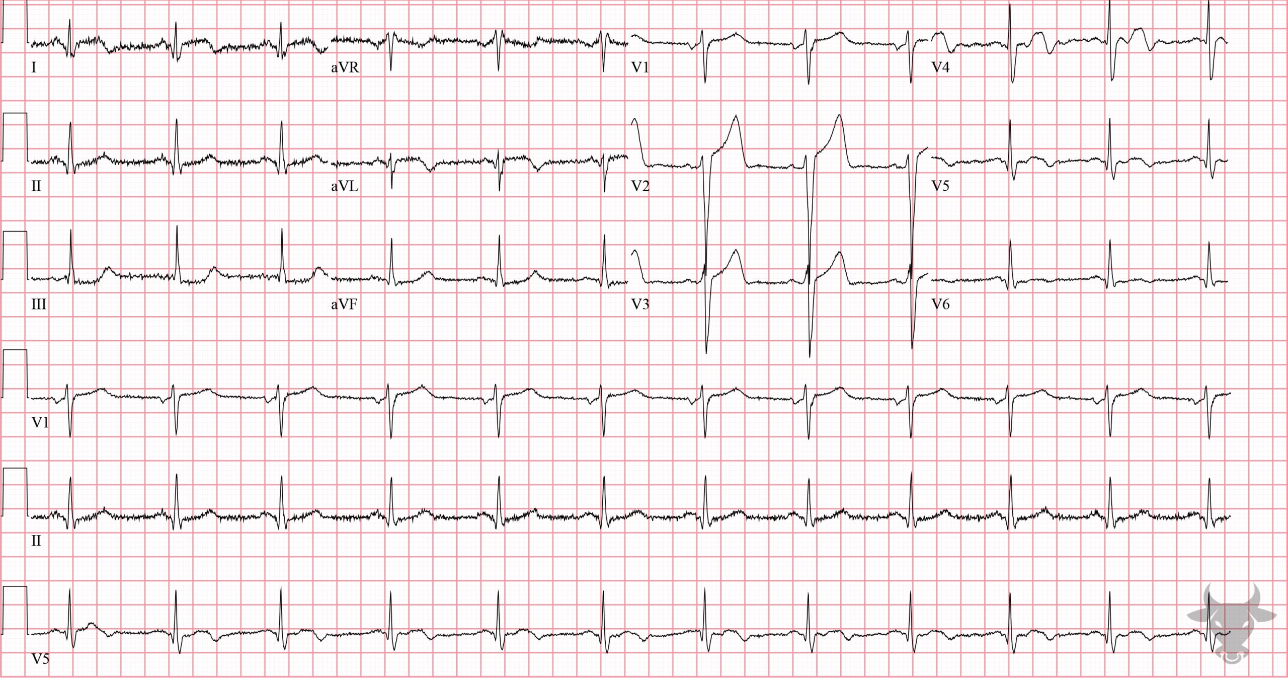
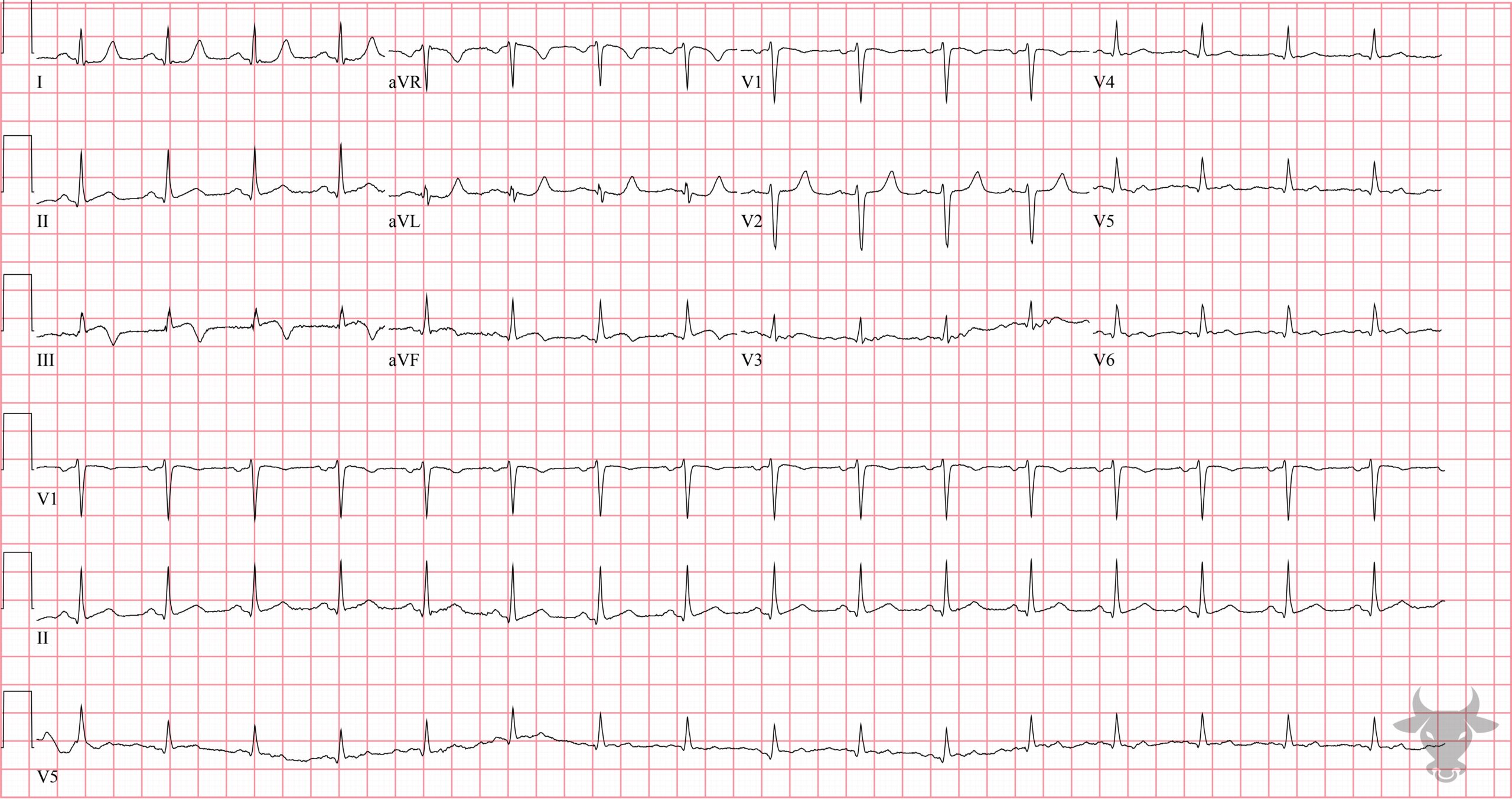
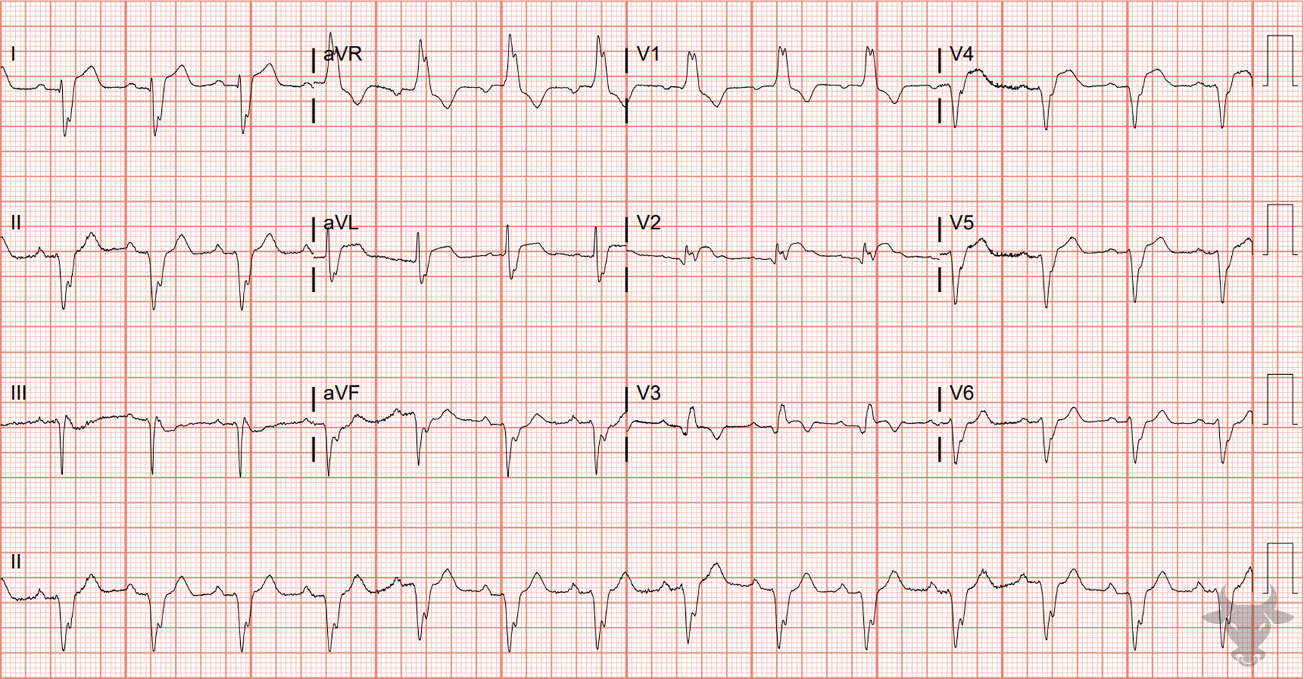
This pattern is eponymously known as the Aslanger pattern. This pattern of isolated ST-segment elevation in lead III, ST-segment depression in V4 through V6, and ST-segment elevation in V1 greater than V2 was first introduced and published in 2020 and is a strong predictor of a culprit plaque in the inferior distribution (left circumflex >> right coronary artery) in patients with concomitant multi-vessel disease. This patient had an occluded graft (from prior bypass) in the right coronary distribution.Anterior ST-elevation Myocardial Infarction
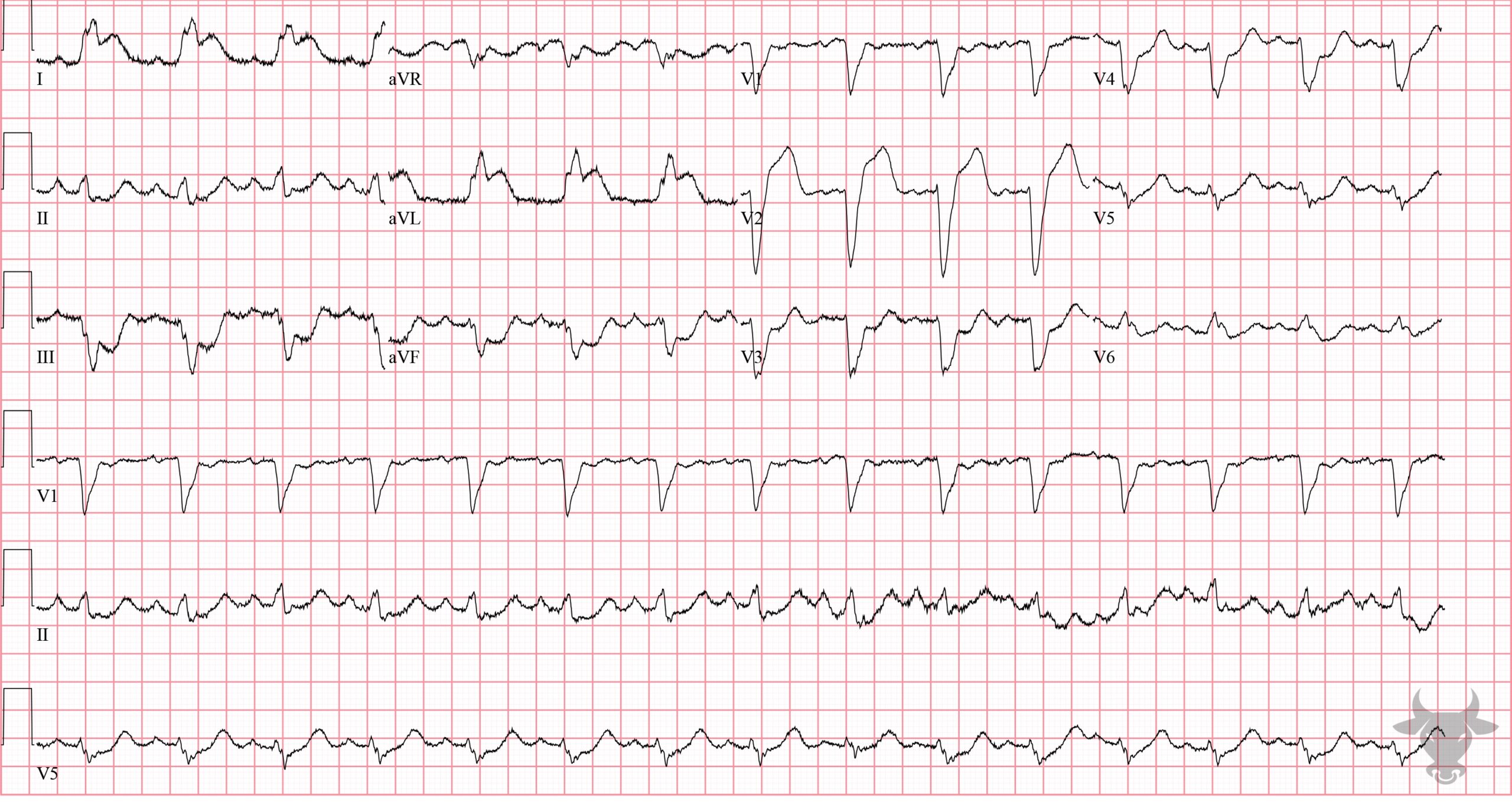
Anterior STEMI. Left heart catheterization showed a distal left main occlusion that was successfully stented.High-lateral ST-elevation Myocardial Infarction
There is subtle ST-segment elevation in the high lateral leads (I, aVL) and subtle reciprocal ST-segment depressions in the inferior leads (II, III, aVF). The T wave in V4 is biphasic with terminal negative deflection and is very specific for ischemia. Later, the T wave in V4 "pseudonormalized" (i.e., became upright) which suggests acute infarct. Left heart catheterization revealed 80% proximal left anterior descending artery stenosis and D2 occlusion.Inferior ST-elevation Myocardial Infarction, Third Degree Atrioventricular Block
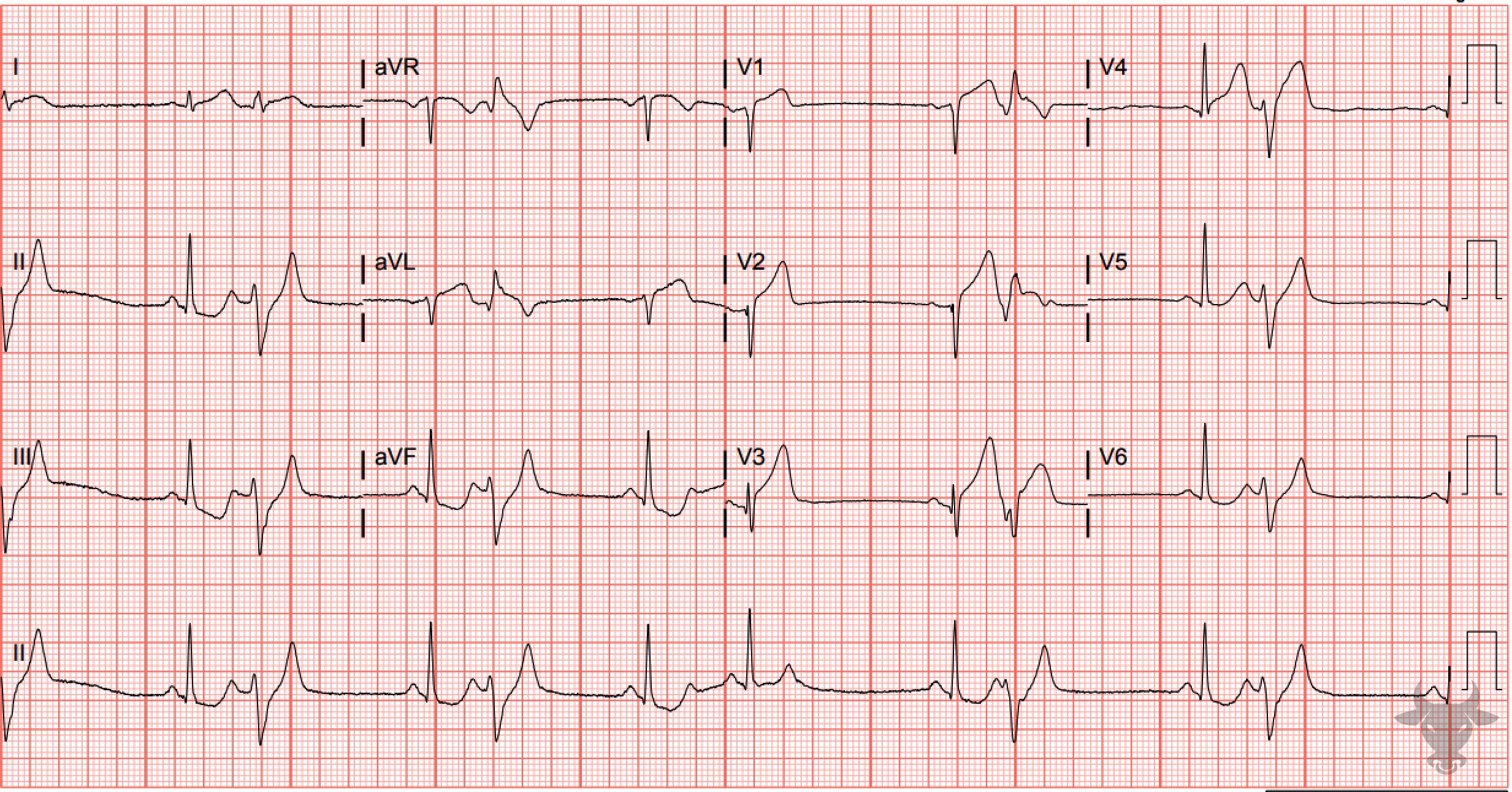
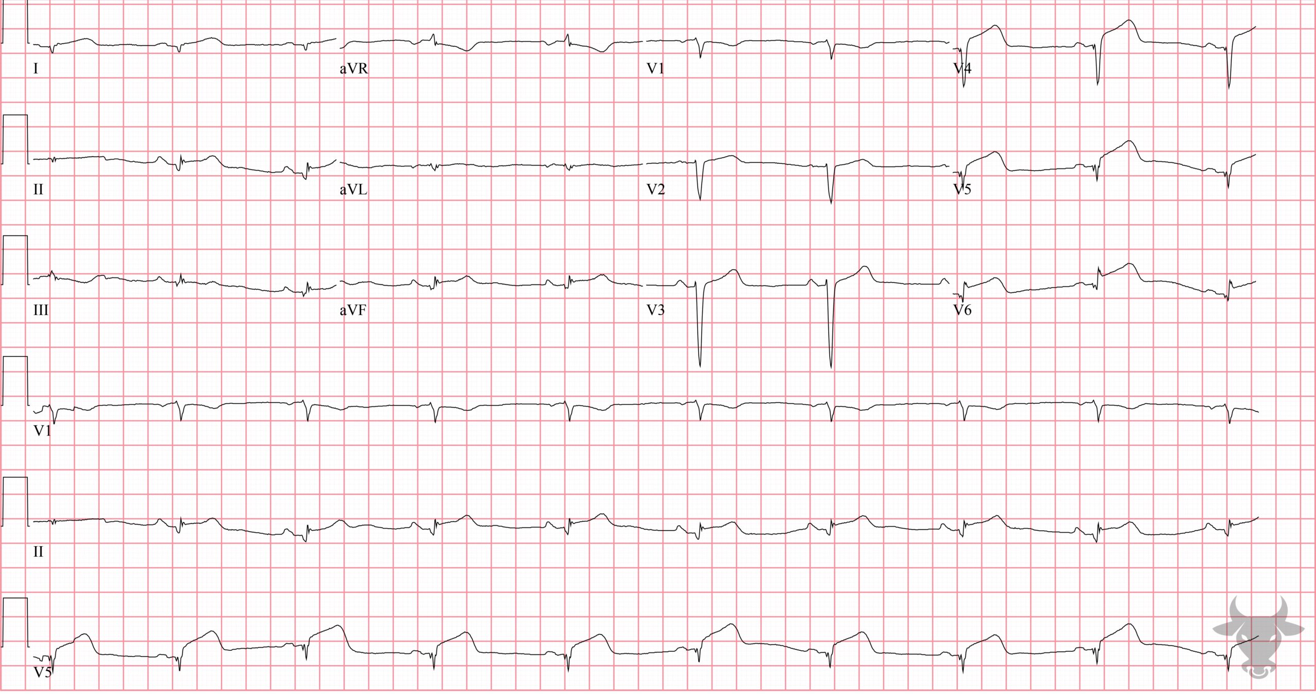
Inferior STEMI with associated complete heart block. The inferior ST segments are “straightened,” and take-off directly from the R wave - an early finding of evolving ST-segment elevation. There are reciprocal ST-segment depressions in aVL and V2. In the setting of inferior STEMI, heart block is usually transient and due to either ischemia or edema of the atrioventricular node, resulting in a junctional escape rhythm. This patient received percutaneous coronary intervention to the right coronary artery after an occlusion was discovered on catheterization.Inferior ST-elevation Myocardial Infarction
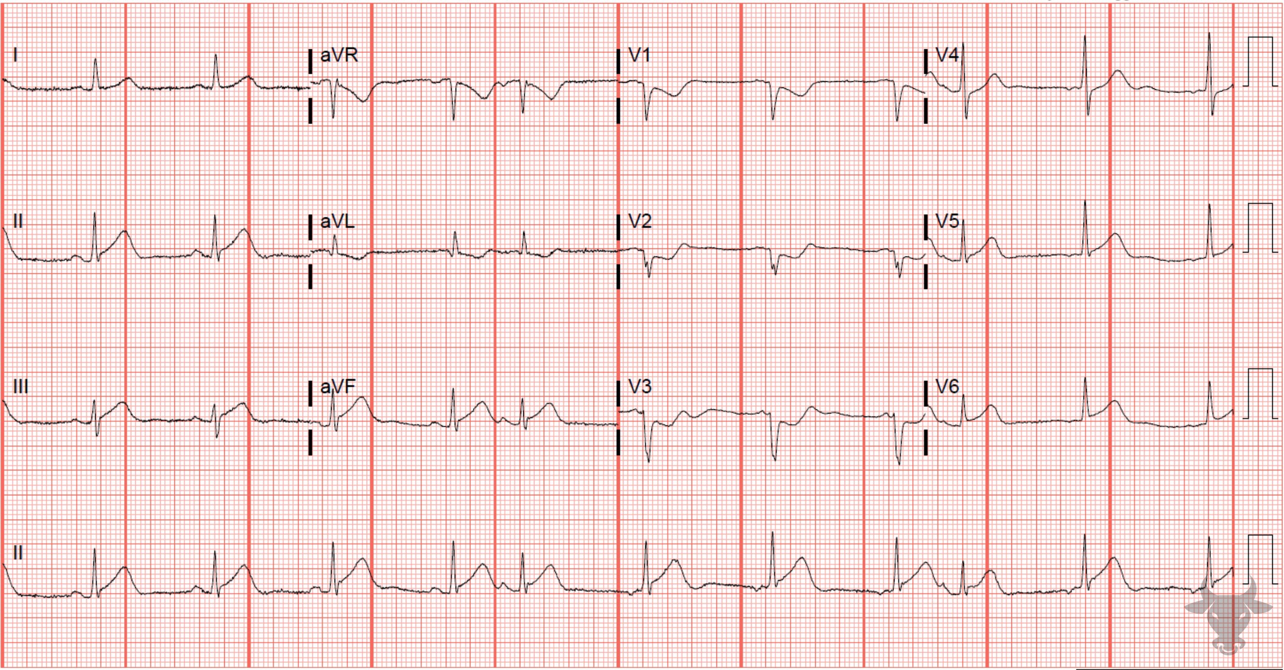
The concave up ST-segment morphology along with the J wave notching might suggest early repolarization; however, the new T wave inversion and slight ST-segment depression in aVL strongly suggests inferior STEMI. This patient received percutaneous intervention to the distal right coronary artery after an occlusion was discovered on left heart catheterization.Anterior ST-elevation Myocardial Infarction
Hyperacute T waves are present in V4 and V5. Note that the amplitudes of the T waves in these leads are greater than the QRS complexes. This patient had a proximal left anterior descending artery occlusion and received percutaneous coronary intervention.Anterior ST-elevation Myocardial Infarction
Anterior STEMI in the setting of a right bundle branch block. Typically, with a right bundle branch block, the ST-segments are slightly depressed in leads V1-3; therefore, any degree of ST-segment elevation in these leads is concerning for anterior STEMI. This patient had a 95% culprit lesion to the left main and received percutaneous coronary intervention.Anterior ST-elevation Myocardial Infarction
The presence of terminal QRS distortion is highly specific for STEMI. Terminal QRS distortion is the absence of an S wave or J wave notching in either V2 or V3. In this case, V3 has terminal QRS distortion. Left heart catheterization revealed a 100% mid left anterior descending artery occlusion.Prior Inferior Myocardial Infarction
Large Q waves in the inferior leads (II, III, aVF) create the left axis deviation. This patient had an old inferior infarction with residual Q waves.Anterior ST-elevation Myocardial Infarction
This patient with an anterior STEMI was confirmed to have a proximal left anterior descending artery occlusion. Inferior ST-elevation Myocardial Infarction
Inferior STEMI with reciprocal changes in aVL. This patient had a proximal right coronary artery occlusion and received percutaneous coronary intervention.Inferior ST-elevation Myocardial Infarction
Subtle inferior ST straightening and concave morphology. Left heart catheterization revealed 90% stenosis of the left circumflex and occlusion of the second obtuse marginal branch.Anterior ST-elevation Myocardial Infarction
Anterior STEMI with concave morphology in V3-5 and poor R wave progression. Anterior STEMIs often do not have reciprocal ST-segment depressions as with this case. This patient received percutaneous coronary intervention of an occluded left anterior descending artery. Inferolateral ST-elevation Myocardial Infarction
Inferolateral STEMI with left circumflex occlusion.Lateral ST-elevation Myocardial Infarction
ST-segment elevation with straightening in V4-6. Left heart catheterization revealed a mid-left anterior descending artery occlusion.Inferior ST-elevation Myocardial Infarction
Subtle inferior ST-segment elevation with anterior precordial reciprocal ST-segment depression. Left heart catheterization revealed a mid-left circumflex occlusion.Hyperacute T Waves
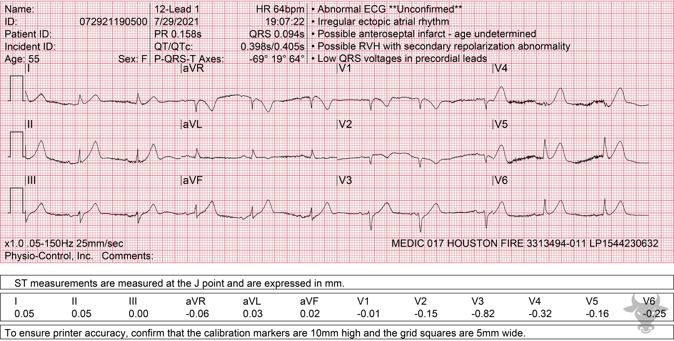
Notice that the amplitude of the T waves exceeds that of the QRS complexes in some leads. This patient had a completely occluded left anterior descending artery.Sgarbossa Criteria – Concordance
Concordant ST-elevation in I and aVL; concordant ST-depression in V3. This patient received percutaneous coronary intervention of an occluded first diagonal branch of the left anterior descending artery.Hyperacute T Waves
Note the hyperacute 'deWinter T waves' that ultimately evolved into an anterior STEMI. Left heart catheterization showed a distal left main occlusion that was successfully stented.Hyperacute T Waves
The T waves in the anterior precordium approach the size of the QRS complexes, making them hyperacute. There are ST depressions in the inferolateral leads. Left heart catheterization revealed an acute left anterior descending artery occlusion.Sgarbossa Criteria – Concordance
Concordant ST-segment elevation in I, aVL in setting of left bundle branch block is specific for acute myocardial infarction. Left heart catheterization revealed diffuse disease and percutaneous intervention was performed to the left anterior descending and left circumflex arteries.Posterior Myocardial Infarction
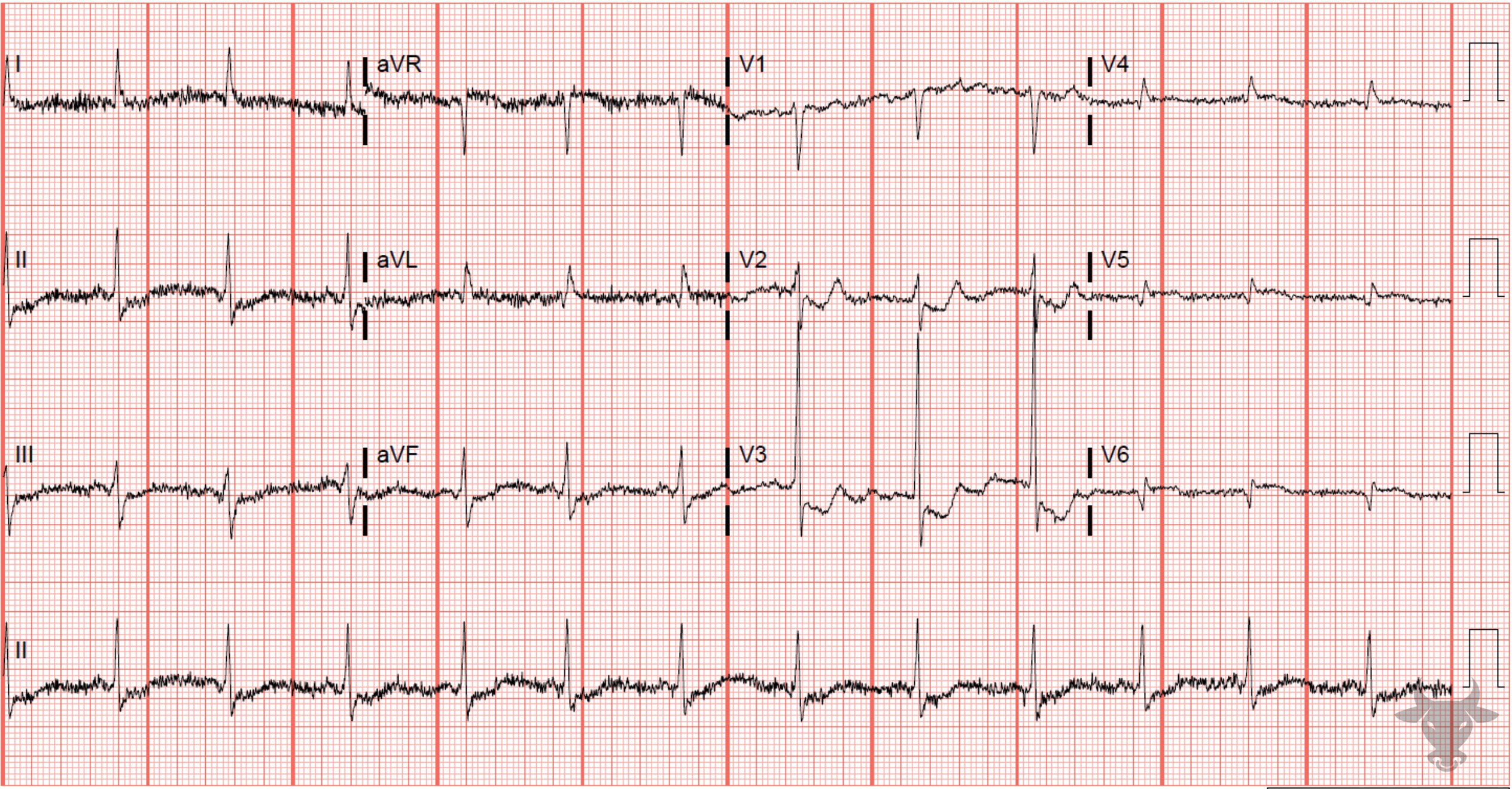
Posterior myocardial infarction with ST-segment depression and upright T waves in the precordial leads V2 through V4. This patient had percutaneous coronary intervention to an occluded left circumflex artery.Posterior Myocardial Infarction
V4-V6 represent posterior leads. At least 0.5mm ST-segment elevation can be seen in V6, confirming the diagnosis of posterior myocardial infarction. This patient had percutaneous coronary intervention to an occluded left circumflex artery.Hyperacute T Waves
While not particularly large at first glance, the T waves in V5 and V6 are larger than the amplitude of the QRS complex and, therefore, represent hyperacute T waves. This ECG evolved into a more obvious STEMI. A completely occluded left anterior descending artery was discovered on left heart catheterization.Hyperacute T Waves
Hyperacute T waves. Left heart catheterization revealed a mid-left anterior descending artery occlusion.Accelerated Idioventricular Rhythm
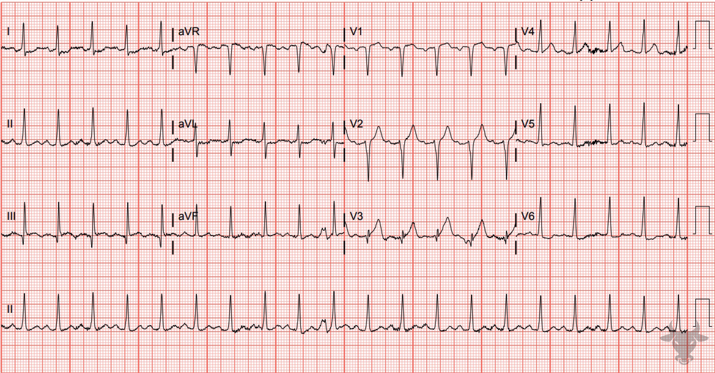
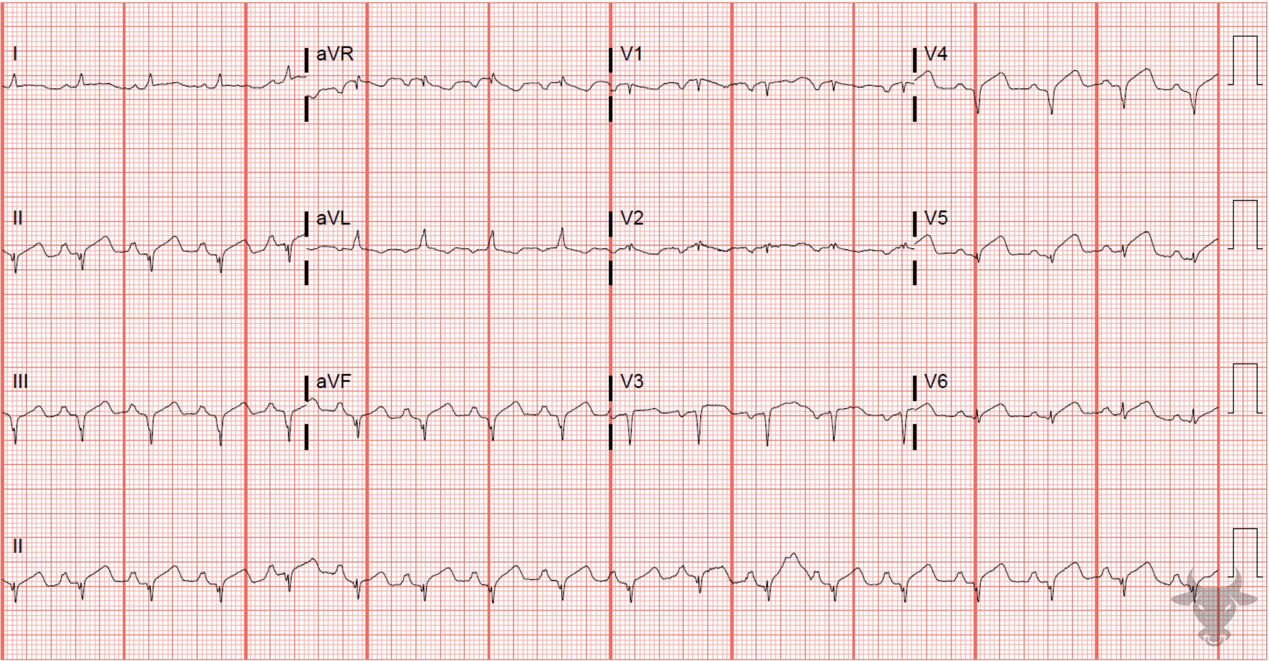
This ECG represented spontaneous reperfusion. The patient was confirmed to have a 90% occluded obtuse marginal culprit lesion. Anterior ST-elevation Myocardial Infarction, Left Ventricular Hypertrophy
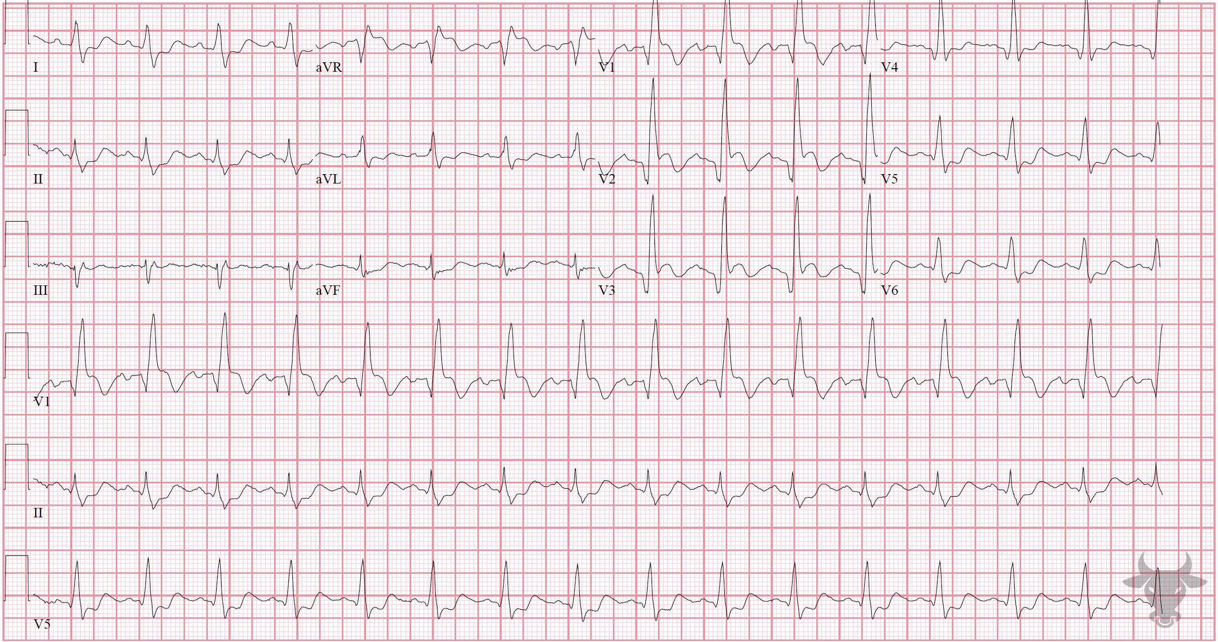
Left ventricular hypertrophy is present given the large QRS complexes. The degree of ST-segment elevation in the anterior precordial leads is more than expected for appropriate discordance. Left heart catheterization revealed complete left anterior descending occlusion.Inferior STEMI With Posterior Extention
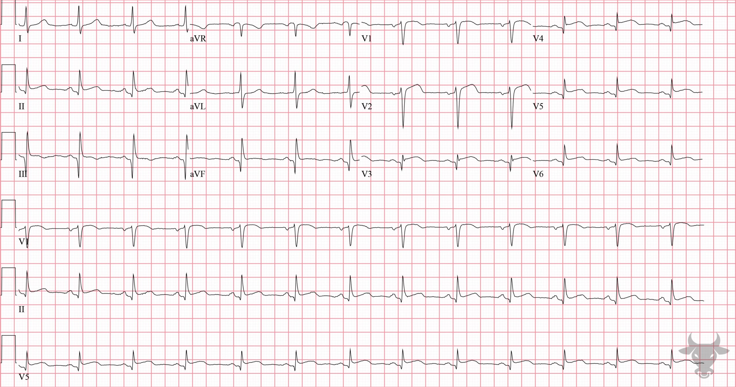
ST-segment elevation with straightening in the inferior leads suggests inferior wall STEMI. The V2 ST-segment depression, large R wave, and upright T wave all suggest posterior extension. Left heart catheterization revealed an occluded distal right coronary artery. ST-elevation Myocardial Infarction With Right Bundle Branch Block
Typically, a right bundle branch block has either isoelectric or slight ST-segment depressions in the anterior precordial leads (V1 through V3). Any degree of ST-segment elevation in V1 through V3 in the setting of a right bundle branch block is worrisome for acute occlusion. This patient had a left anterior descending artery occlusion. 